![]()
![]()
![]()
Yongyut Predalumpaburt
Seabass spawn naturally in captivity during the hours of 2000 to 2100. Fertilized eggs need 12 to 15 hours in 29.5–31 degrees Celsius seawater for hatching. The spherical eggs range from 0.74 to 0.80 cm in diameter with a single oil globule from 0.20–0.28 mm in diameter (Maneewong and Watanabe, 1984).
Details of larvae and juvenile development based on the samples from hatchery rearing at the National Institute of Coastal Aquaculture (NICA) are as follows:
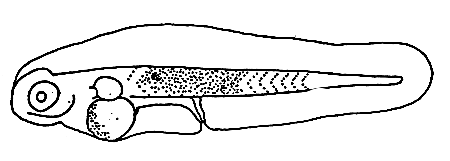
1. Larva at 2.05 mm (1 day after hatching)
Yolk absorbed. The single oil globule at the anterior end of yolk. Mouth still closed. Anus situated near the middle of body. Eyes unpigmented. Pectoral fin appeared as bud.
2. Larvae at 2.75 NL. mm (4 days after hatching)
The mouth opened with differentiated upper and lower jaws. The nostrils located on each side of snout. The pectoral fin developed in round shape as finfold. The digestive tract extended relatively in thickness. Melanophores present on many parts of body. Stellate and blotch melanophores scattered on mid—brain, lower jaw angle. The row of branch melanophores occurred on dorsal contour, ventral colour, body mid—line, the smaller ones on ventral of abdomen.
* Senior Fishery Biologist, NICA.
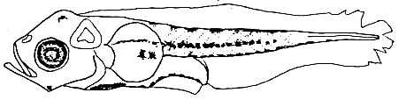
3. Larvae at 3.60 NL. mm (10 days after hatching)
The larvae developed three prepercular spines on preopercle, The head slightly round and the body depth extended with developing dorsal and anal fin base. The tip of notochord just flexed to develop the caudal fin. Melanophores increased densely on various parts of the body such as dorsal contour, ventral contour, body mid—line and also appeared on nasal, lower jaw angle, preopercule, dorsal and ventral of digsetive tract, caudal fin base.
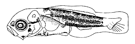
4. Larvae at 3.80 NL. mm (13 days after hatching)
The number of preopercular spines increased to five and the spines on angle of preopercule was slightly bigger than the others. The nostrils still not separate. The gut fully extended. The dorsal and anal fin were developing with increasing fin ray. The medial fin fold, behind the dorsal and anal fin were separated to form the dorsal and anal fin. The caudal fin was round and completely developed fin rays.
The melanophores increased gradually on the tip of snout, lower jaw, otic capsule and body. The blotch melanophores on body such as dorsal and ventral contous, body mid—line extended to form three dark bands on body.
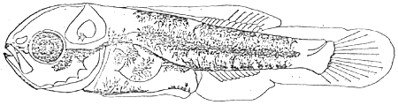
5. Juveniles at 5.5. mm. SL (18 days after hatching)
The nostrils completely separated in two pores. Maxillary reached to the center of eye. The single opercular spine presented on the upper part of opercule. Body depth expanded relatively. The dorsal, anal and caudal fin completely developed spines and rays as follows: D.VIII, 8; A III, 8; C6, 5. The pectoral fin was developing fin ray while the pelvic fin appear as bud. The number of preopercular spines increased to six but its size was relatively small. The blotch malanophores scattered on head and most of body part and the posterior part of body also remained branched melanophores on dorsal and ventral as well as body mid—line. The body has two vertical bands and divided by mid—point of body. There was no melanophore on caudal peduncle.
6. Formation of fin
The seabass larva formed dorsal, anal and caudal fin when the total length reached 3–4 mm or at 7 days after hatching.
The first seven spines of dorsal fin were formed at 6 mm TL. The eighth spine still liked a soft ray and the spine became a, spine at 8–12 mm TL. So the number of fin rays and spine became constant (VIII, 11) when the larvae reached 8–12 mm TL. The segmentation of soft dorsal fin was started from 5 mm TL and then completed at 6–16 mm in TL as well as the branching of soft dorsal fins was started from 20 mm TL but was still not completed at 44 mm TL.
The first two spines of anal fin were formed at 6 mm TL while the third one was still like a soft ray. After the larvae reached the size 8–11 mm TL, the anal fin was completely developed and its formula was A III, 8. The segmentation of anal fin was the same as the dorsal fin but branching was completed at 34–38 mm TL.
The pelvic fin developed rapidly from 5 mm TL and completed at 8 mm TL. The formula was P I, 5. The segmentation of pelvic fin was started at 12–16 mm TL.
The caudal fin developed after the notochord flexed. The total number of fin ray which contained rudimentary soft rays became constant (30–33) at 11–12 mm TL. The segmentation of the caudal fin was started from 3–4 mm TL and completed at 12–15 mm TL. Btanding was started from 12–17 mm TL and completed at 31–35 mm TL.
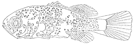
The larvae of the seabass became juveniles when the total length reached 8–12 mm or 20 days after hatching. Furthermore, branching of soft ray in all fins except pectoral fin might be completed before 35–50 mm TL so that it seems that the juvenile may become the young fish at that size.
REFERENCES
Kosutarak, P. and T. Watanabe. Notes on the development of larval and juvenile stages of seabass. p. 36–45. Report of Thailand and Japan Joint Coastal Aquaculture Research Project. No. 1. JICA. FDT. 84-35. 189p.
Maneewong, S. and T. Watanabe. 1984. Record of spawning and hatching of egg of seabass, Lates calcarfier, in concrete tank. p. 46–57. Report of Thailand and Japan Joint Coastal Aquaculture Research Project. No. 1. JICA. FDT 84-35. 189p.
![]()
![]()
![]()