Dropsy of Mixed Aetiology in Catla catla (Ham.)
| NACA/WP/86/31 | November 1986 |
|
Dropsy of Mixed Aetiology in Catla catla (Ham.) |
Central Institute of Freshwater Aquaculture
Dhauli, Kausalyagang, Bhubaneshwar
NETWORK OF AQUACULTURE CENTRES IN ASIA
BANGKOK, THAILAND
Hyperlinks to non-FAO Internet sites do not imply any official endorsement of or responsibility for the opinions, ideas, data or products presented at these locations, or guarantee the validity of the information provided. The sole purpose of links to non-FAO sites is to indicate further information available on related topics.
This electronic document has been scanned using optical character recognition (OCR) software. FAO declines all responsibility for any discrepancies that may exist between the present document and its original printed version.
Dilip Kumar, B.K. Mishra and R.K. Dey
Unit for Ichthyopathology and Fish Health Protection
Freshwater Aquaculture Research and Training Centre
(CIFRI)
P.O. Kausalyagang, Via Bhubaneshwar (Orissa) 751002
INDIA
Dropsy in Catla catla (Ham.) involving mixed infection of protozoan parasite and a bacterium is described. Aetiological agents of the disease - myxosporidian parasite and a strain of Aeromonas hydrophila have been isolated and identified. External clinical symptoms together with pathoanatomical and histopathological changes are presented and important implications of the mixed infection discussed towards understanding the disease process.
INTRODUCTION
Like other intensive aquaculture systems, composite fish culture also aims at increasing fish production by various intensification approaches such as increased stocking rates, increased feeding and fertilization programme. Under such conditions it is inevitable that imminical conditions enhancing the development of disease will be encountered (Kumar et al., 19 ) Several workers have already reported occurrance of diseases in composite fish culture ponds (Pal and Tripathi, 1978; Dey et al., 1982). During routine examination of fish samples from composite fish culture ponds under fish health monitoring programme, authors came across few cases of dropsy in Catla catla at several occasions. Thorough investigations into dropsy disease in fish dates back to 1930 by Schaperclaus. Infectious Dropsy and Hydrops have also been described in European carp farms (Tomasec, 1941; Otta, 1963 and Ghittino, 1968). Gopalakrishnan (1961) has reported infectious dropsy of Indian major carps and its experimental induction. Most authors considered infectious Dropsy of carp (IDC) to be contagious, but expressed doubt about its aetiology. Some workers considered it to be a bacterial, some a viral and others a disease caused by a combination of both virus and bacterium. Only recently Fijan and his associates have isolated and named a virus Rhabdoyirus carpio as the most likely cause of acute infectious dropsy of carps (Fijan, 1972). However, the etiological agent of the infections dropsy in cultured major carps in India has been reported to be species of Aeromonas (Gopalakrishnan, 1961). Pal and Tripathi (1968), though did not isolate the casual agent, could treat the disease fully using terramycin in the feed. However, the present cases are interesting from the view of its mixed aetiology from which strain of bacterium Aeromonas hydrophila ssp. hydrophila and spores of myxosporidian parasite have been isolated. The present observation aims towards understanding the disease process.
Paper presented in the Fourth All India Seminar on Ichthyology,
Department of Zoology, D.A.V. College, Dehra Dun, India in
September 1983.
MATERIALS AND METHODS
Specimens of catla (Catla catla) with signs of dropsy were collected at two occasions from nearby composite fish culture pond adopted under ‘Lab-to-Land Programme’ and also from reservoir-I of the Centre during the course of routine sampling for fish health monitoring. All the 5 diseased specimens were of 1+ age having weight range of 400 g to 500 g., while other healthy specimens of the same stock grew over 1 kg. Sampling were closely examined for external clinical symptoms and swabs were taken with full aseptic precautions from kidney and heart for isolation and study of bacterial pathogen using diagostic schem of Shotts ans Bullock (1975). Later on, the strains were compared with Hungarian referent strain H-604 (Aeromonas hydrophila ssp. hydrophila) (Kumar et al., 1985). Fish were then dissected for pathoanatomical observations. Fluid smear from abdominal and swim bladder cavity and squash preparations of kidney, liver, gills and spleen etc. ere examined immediately. Samples were also taken for detailed histopahtological and parasitiological examinations. Myxosporidian spores were isolated from kidney using trypsin digestion technique.
RESULTS
All the specimens were highly emaciated with distended abdomen and compared to healthy ones deeply pigmented. Epidermis sloughed off towards posterior region of the head, scales were raised with watery fluid filled in scale pockets, superficial ulcerations and haemorrhage on the skin at some places of the body (Fig. 1). Skin portion of the opercular flap was also found onlarged a bit. Laboratory examinations revealed emaciation, anaemia, accumulation of slight opaque fluid in the skin and the abdominal cavity, haemorrhages in the skin and pale gills. The liver was dark brown in colour. Intestine was empty with yellow mucous. Incision of the kidney resulted in the semi-fluid contents dripping out. In one such specimen posterior chamber of the swim bladder was found collapsed, twisted and thickened and had a narrow bore with little air and fluid in. Squash preparations of the kidney of infected carps showed the presence of cysts and numerous myxosporidian spores with various developmental stages. Kidney impressions stained with Gram's stain showed the presence of myxosporidian spores as well as numerous rod shaped gram-negative bacteria. Gram stained smear preparations of fluid from abdominal and swim bladder cavities demonstrated presence of myxospiridian spores and gram-negative rods. Excessive pigment accumulation was found in the liver parenchyma causing extensive damage due to pigment infilteration throughout the liver madd. Marked chordal disarray was also noticed. Myxosporidian cysts showed its presence in kidney sections and degeneration and necrosis of tubular epithelium was also prominent (Fig. 2). Areas of haemorrhage showing inflamatory process in the kidney tubules were also observed. Marked epithelial proliferations was found in the posterior chamber of the swim bladder and at some points the epithelium has thickened and has become multilayered in one specimen.
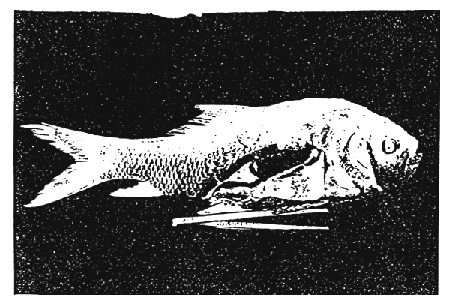
Fig. 1 Pathomorphology and pathoanatomy of diseased specimen of Catla catla
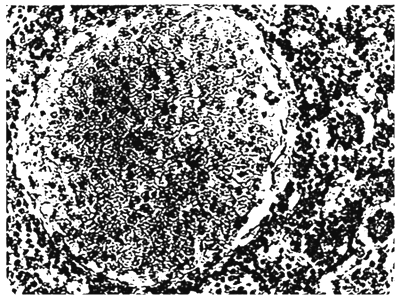
Fig. 2 Myxosporidian cyst in kidney section
Swabs taken from kidney of diseased specimens produced yellow colonies on Rimler-Shotts medium (Shotts and Rimler, 1973). Pure isolates of the dominant form were found gram-negative, cytochrome oxidase positive, fermentative, respiratory and presumptively identified as strains of Aeromonas hydrophila. These isolates were similar to Hungarian referent strain H-604 of Aeromonas hydrophila ssp. hydrophila, though the butanediol test was always negative.
DISCUSSION
There are very few truly obligate bacterial pathogens of warm water fish. Most bacterial diseases noted are secondary to some 'stress' placed upon the fish. Primary offenders producing stressful conditions include, various water quality parameters and/or parasitic infestations (Bullock et al., 1971 and Shotts, 1979). Wedemeyer and Wood (1974) have also discussed the stress mediated diseases in Pacific salmon, trout, catfish, carpand shad. Bererano et al. (1979) have reported mass mortalities in silver carp associated with bacterial infection following halndling. Walters and Plumb (1980) have also correlated stress and bacterial infection in Channel catfish and Kumar et al. (1982) have reported stress mediated columnaris disease in rohu (Labeo rohita) in India. In the present study of dropsyit seems that bacterial infection has most probably occurred in the premortal stage. Myxosporidian infection of the kidney can cause emeciation and anaemia but not the mass appearance of pigment cells and extensive damage in the liver. On the other hand Aeromonas hydrophila can neither cause emaciation nor induce pigment accumulation but can be associated with bleeding (Fijan, -1983). Walters and Plumb (1980) have reported necrosis and oedema in the liver in experimentally infected channel catfish, Ictalurus punctatus by Aeromonas hydrophila under environmental stressors such as low D.O., added CO2 and NH3, while no histopathology was observed in experimentally infected fishes which were not subjected to multiple stressors and were able to eliminate the bacteria. Enzootic nature of renal myxosporidiosis in Indian major carps was ascertained in the districts of Cuttack and Puri of Orissa including the ponds from where samples have been collected on the basis of data collected under Fish Health Monitoring Programme of the Centre (Mishra et al, 1982). Necrotic and degenerative changes in most of the kidney tubules with vacuolar degeneration of cytoplasm and pyknotic nuclei have been observed in reneal myxosporidiosis in such cases. Walters and Plumb (1980) have also found haemorrhage enlargement of tubules and necrosis in kidney due to Aeromonas hydrophila infection. Mistopathological changes observed in these specimens can, therefore, be regarded as concomitant. Histopathological changes observed here in swim bladder have been found to be associated with IDC. Few authors have also considered it to be a symptom of IDC or to be caused by Aeromonas. In the present observation the pathological condition of the swim bladder seems to be associated with dropsy while myxosporidians might have invaded during aggravated health condition of the fish. However, Bachman and Ahne (1973; 1974) isolated a virus indistinguishable from SVC virus from affected fish. According to Roberts (1978) carp dropsy complex and swim bladder inflemmation are still in the process of elucidation. Myxosporidian infection might have stressed the fish sufficiently to predispose them to infection by Aeromonas hydrophila. Malevitskaya (1959) also reported that malignant myxosporidial anaemia of carp may be accompanied during spring by IDC and coccidial enteritis giving a very confused clinical picture.
ACKNOWLEDGEMENT
We are indebted to Dr. A.V. Natarajan, Director, Central Inland Fisheries Research Institute, for his keen interest in the work and Dr. V.R.P. Sinha, National Project Director, FAO/UNDP Projects and Head, Freshwater Aquaculture Research and Training Centre for valuable suggestions and encouragement and also to Prof. Dr. Nilola Fijan, FAO/UNDP Consultant for his helpful guidance.
REFERENCES
Bachman, P.A. and W. Ahne, 1973. Isolation and characterization of agent causing swimbladder inflamation in carp. Nature, Lond., 244 : 235–237.
Bachman, P.A. and W. Ahne, 1974. Biological properities and identification of the agent causing sim-bladder inflamation in carp. Arch. ges. Virusforach., 44 : 261–269.
Bejerano, Y., S. Sarig, M.T. Morne and R.J. Roberts, 1979. Mass mortality in silver carp Hypophthalmichthys molitrix (val.) associated with bacterial infection following handling. Journal of Fish Diseases. 2 : 49–56.
Bullock, G.L., D.A. Conroy and S.F. Sniesmko, 1971. Bacterial diseases of fishes, In: Diseases of Fishes, ed. S.F. Snieszko and J.R. Axelrod, Nepture, N.J. : TFH P. blishers.
Dey, F.K., Dilip Kumar, B.K. Mishra and K. Suresh, 1982. Sampling methods, packing and shipments of materials for laboratory diagnosis of fish diseases. Souvenir: Workshop on the development of Inland Fisheries in Orissa through Institutional Finance FFDA, Balasore (Orissa). 6–8 March.
Fijan, N.N., Z. Petrinee, D. Sulimanovic and L.O. Zwillenberg, 1971. Isolation of the viral causative agent from acute form in infectious dropsy of carp. Vet. archiv, 41 : 125–138.
Fijan, N.N., 1972. Infectious dropsy in carp - a disease complex. In : Diseases of Fish, Proc., Symp. No. 30, Zoological Society, London, May 1971, ed. L.E. Mandesley-Thomas, pp 39–51. New York and London; Academic Press and the Zoological Society.
Fijan, N.N., 1983. Diagnostic work and Research on Fish Diseases and Fish Health Monitoring at FARTC (CIFRI), Dhauli. FX : DP/IND/75/ 031 FAO Field Document 5.
Ghittino, P., 1968. Les maladies contagienses des poissons incluses dans le Code Zooanitaire International de L'O.I.E. Paris : Office International des Epizooties.
Gopalakrishnan, V., 1961. Observations on infectious dropsy of Indian carps and its experimental induction. J. Sci. Industr. Res. (C)., 20 : 357–358.
Kumar Dilip, B.K. Mishra, K. Suresh and R.K. Dev, 1982. Role of Prophylaxis in Aquaculture. Souvenir: Workshop on the Development of Inland Fisheries in Orissa through Institutional Finance, FFDA, Balasore (Orissa). 6–8 March.
Kumar Dilip, K. Suresh, R.K. Dey and B.K. Mishra, 1982. Stress mediated columnaris disease in Rohu (Labeo rohita). Abstract. Symp. Diseases of Finfish and Shellfish. College of Fisheries Mangalore. 1–3 March.
Kumar, D., T. Farkas and V.R.P. Sinha, 1985. Bacteria from diseased fishes in India. Aquaculture Hungarica (Szarvas), Vol. IV, pp.
Malevitskaya, M.A., 1959. Kvoprosu O “Smeshanny - kh” Zabolevaniyakh karpa V ryborodnykh khosyaistvakh (On “Mixed” Diseases of carp in fish Farms) - Trudy Soveshchanica Ikhtiologicheskoi Kommssii AN BSSR, Vol. 9. 1959.
Mishra, B.K., R.K. Dey, Dilip Kumar and K. Suresh, 1982. Observations on renal myxosporidiosis in Indian major carps. Abstract. Proc. Symp. Diseases of Finfish and Shellfish. College of Fisheries, Mangalore. 1–3 March.
Otta E., 1963. Die heutige Ansichten iiber die “Atiologie der” Intektiosen Bauchwassersucht” der karpfen. Wien. tierargtl, Machr., 50 : 995–1005.
Pal. R.N. and S.D. Tripathi, 1978. J. Inland Fish. Soc. Ind. Vol. 10; 166–168.
Richards, R.H. and R.J. Roberts, 1978. The Bacteriology of Teleosts. In: Fish Pathology. ed. R.J. Roberts. Bailliere Trinidall. London Publishers.
Schaperclaus, W., 1930. ‘Pseudomonas puntata als’ Krankheitserreger bei Fischen'. Zeitschrift fur Fishcherei. XXVIII, No. 3 : 290–370
Shotts, E.B., 1981. Bacterial Diseases of Fish. Proc. Republic of China - United States Cooperative Science Seminar on Fish Diseases. NSC Symposium series No. 3, National Science Council, Republic of China.
Shotts, E.B. and G.L. Bullock, 1975. Bacterial Diseases of Fishes. Diagnostic Procedures for Gram-Negative Pathogens. J. Fish. Res. Bd. Can., 32 : 1243–1247.
Shotts, E.B. and R. Rimler, 1973. Medium for the Isolation of Aeromonas hydrophila. Appl. Microbiology, Oc., 1973. : 550–553.
Tomasec, I., 1941. O biti t. zv. zarzne vodene bolesti (Pdrudomonas infekcje) Sarana. Vet. archiv., 11 : 257–278.
Wedemeyer, G.A. and J.W., Wood, 1974. Stress as a predisposing factor in fish diseases. U.S. Fish. & Wildl. Sev., FDL 38. 8 pp.

![]()