![]()
![]()
![]()
Plates 30 & 31 (pp. 211–212)
All data are on individual fish picked up from collected samples or commercial catches due to their obvious malformation, anorexia or distinct tumour-like growths. Additionally there is a fair number of documented descriptions of neoplasia in aquarium reared African fish. Some of these conditions are worth elaboration, others will be briefly mentioned. Malformations occurring in cultured fish or induced following hybridisations are not included due to their epizootiological irrelevance. The same applies to congenital anomalies reported from cultured stock (Roberts and Sommerville, 1982). Neoplasia and degenerative chronic conditions are often linked to anthropogenic stresses on the ecosystem, in particular due to release of xenobiotic materials to the environment (Edwards & Overstreet, 1976; Hawkins et al., 1988). These situations are less obvious in the less developed regions and, although in some places they may exist, relevant documentation is presently lacking.
20.1.1 ANOREXIA WITH METAPLASIA AND NEOPLASIA in Clarias gariepinus in Lake Victoria (Plate 30a-c)
Emaciated male and female specimens, both over 70 cm long. Necropsy of the female revealed subdermal myxofibroma (or myxofibrosarcoma), grossly enlarged spleen due to haemangioma (or haemangioendothelioma) and the ovary comprised of several large (up to 4 cm diameter) cysts full of serous liquid. In the male fish only the testes were slightly enlarged with seminiferous tissues replaced intermittently with zones of fibrosis and necrosis (Tumour identification by Dr J.C. Harshbarger, Registry of Tumor in Lower Animals, Smithsonian Museum, US Nat. Mus.- accession no. RTLA 759A & B).
20.1.2. EMACIATED Protopterus aethiopicus FROM L. VICTORIA, with no otherwise detectable gross pathology
20.1.3. EPITHELIOMA IN TWO FULL SIZED Malapterurus electricus from L. Albert. (Plate 31 a-e)
Tumours occurred on the lips, head and flanks. Proliferative tissue was of two types, one involving predominantly regular skin epithelial cells (malpigi cells) and the other consisting mainly of the glandular (albuminoid) "fear cells" (RTLA 760; Paperna, 1975).
20.1.4 Haplochromis sp. FROM L. VICTORIA with subcutaneous lipoma (RTLA 761) Plate 31g (p. 212)
20.1.5 MATURE Synodontis afrofisheri WITH CURVED SPINE from L. Victoria (Plate 30 d-p. 211)
Haller & Roberts (1980) report a case of dual neoplasia in a specimen of Sarotherodon spilurus from brood stock held in a brackish water farm near Mombasa, Kenya. A large tumour on the flank consisted entirely of lymphocytes-a lymphoma, and the second in the kidney was identified as a renal tubular adenoma.
A case of multiple fibroma has been reported from Malapterurus electricus and two cases of a spermatocytoma, from Protopterus aethiopicus in the USA and Japan; the former also with renal melanoma (Stolk 1957a; Nigrelli & Jakowska, 1953; Prince Masahito et al., 1984). Adenoma of the pharyngeal glands was described in Haplochromis multicolor (Stolk, 1957b) and Osteochondroma in Hemichromis bimaculatus (Nigrelli & Gordon, 1946).
REFERENCES
Edwards, R.H. & Overstreet, R.M., 1976. Mesenchymal tumors of some estuarine fishes of the northern gulf of Mexico. I.Subcutaneous tumors, probably fibrosarcomas, in the striped mullet, Mugil cephalus. Bull.Mar.Sci., 26: 33–40.
Haller, R.D. & Roberts, R.J., 1980. Dual neoplasia in a specimen of Sarotherodon spilurus spilurus (Gunter) (=Tilapia spilurus). J. Fish Dis. 3: 63–66.
Hawkins, W.E., Overstreet, R.M. & Walker, W.W., 1988. Small fish models for identifying carcinogens in the aqueous environment. Water Resour. Bull., 24: 941–949.
Nigrelli, R.F. & Gordon, M., 1946. Spontaneous neoplasms in fishes. 1. Osteochondroma in the jewel fish, Hemichromis bimaculata. Zoologica N.Y., 31: 89–92.
Nigrelli, R.F. & Jakowska, S.J., 1953. Spontaneous neoplasms in fishes. VII. A spermocytoma and renal melanoma in an African lung fish Protopterus annectens (Owen). Zoologica N.Y. 38: 109–112.
Paperna, I., 1975. Skin epithelioma in the electric catfish Malapterurus electricus from L. Albert, East Africa. Copeia, 1975 (2): 374–378.
Prince Masahito, T., Ishikawa, T. & Takayama, S., 1984. Spontaneous spermatocytic seminoma in African lungfish, Protopterus aethiopicus Heckel. J. Fish Dis., 7: 169–172.
Stolk, A., 1957a. Tumors of fishes. 13. Multiple finromas of the skin in the malapterurid Malapterurus electricus. Proc.K.Ned.Akad.Wet. (C.Biol.Med.Sci.), 60: 41–52.
Stolk, A., 1957b. Tumors of fishes. 17. Adenoma of the pharyngeal gland. Proc.K.Ned. Akad.Wet. (C.Biol.Med.Sci.), 60: 640–649.
ILLUSTRATIONS
Plate 30. Deformations, degenerations and neoplasia: (p. 211) a. Emaciated Clarias gariepinus, from L. Victoria. Necropsy of such fish revealed neoplastic processes as well as degenerative changes in the gonads (see b,c,f). b. Degenerative testes with parts of the seminiferous tissue being replaced by connective tissue and necrotic deposits. c. Cystic ovary of specimen a. d. Spinal curvature in adult Synodontis afrofisheri from northern L. Victoria (x 0.6).
Plate 31. Neoplasia and other abnormalities (p. 212 with legend).
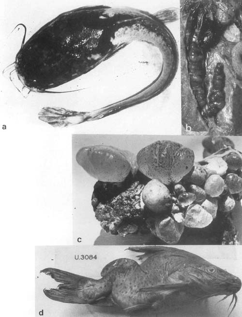
Plate.30. Deformations, degenerations and neoplasia. (Legend p.210)
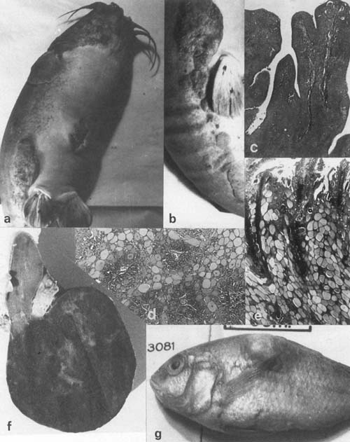
Plate 31. Neoplasia and other abnormalities; a–e. Epithelioma in Malapterurus electricus from northern L. Victoria. a,b. Gross pathology. Histology of proliferative epithelium from the mouth region, where only malpigi cells are involved. c–d. Skin area where glandular, albuminoid cells are also involved. f. Spleen with large tumour identified as haemangioendothelioma. g. Haplochromis sp. with large dermal lipoma.
![]()
![]()
![]()