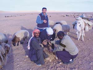
Active surveillance of RVF was carried out through the monitoring of sentinel herds |
Serological and clinical data generated for the third consecutive year by the FAO-supported regional Rift Valley fever (RVF) surveillance system contributed to a better understanding of the epidemiology of the disease at the regional level, as well as countries' enhanced preparation to respond to occurrences of the disease. Active surveillance of RVF was carried out through the monitoring of sentinel herds (sera were collected from 785 small ruminants and tested for the detection of IgM and IgG antibodies) from July to November 2002 in Mauritania and Senegal.
 |
RVF outbreaks in Galoya (Matam) and Thilogne (Podor) |
 |
RVF risk map: potential areas of risk in Western Africa |
In Mauritania, viral circulation was observed in August in the Assaba region, Keikratt (four IgM-positive sheep and goats out of 30), and a human case was detected in November (village of Bachaat, Mbout, Gorgol region). Two clinical suspicions were also investigated, but were found to be negative in sero-neutralization.
In Senegal, there was no evidence of viral circulation in September and October (no antibodies were detected by serological monitoring). However, at the end of the rainy season, frequent abortions were reported in Galoya and Thilogne, and a mission was sent to conduct an epidemiological investigation. In total, the disease was suspected on four occasions, and two outbreaks were confirmed.
Serological results (IgM ELISA) in small ruminants displaying clinical signs compatible with RVF |
|||||||
No. |
Village |
Type of |
Species |
Sex |
Age |
Clinical signs |
ELISA IgM* |
1 |
Bile |
Serum |
Ovine (mbortou ndiamalou) |
Female |
1 year |
Abortion |
1.137 |
2 |
Bile |
Serum |
Ovine (hérou ndiamalou) |
Female |
3 years |
Abortion |
1.397 |
3 |
Bile |
Serum |
Ovine (powro) |
Female |
1 year 6 months |
Abortion |
1.397 |
4 |
Bile |
Serum |
Ovine (ndakou danedji) |
Female |
2 years 6 months |
Abortion |
1.366 |
5 |
Bile |
Serum |
Ovine (hirqué danedio) |
Female |
2 years 6 months |
Abortion |
1.215 |
6 |
Bile |
Serum |
Ovine (hirquewou mboulou) |
Female |
2 years |
Abortion |
1.392 |
7 |
Bile |
Serum |
Ovine (ndakou ndiadou) |
Female |
2 years |
Abortion |
1.357 |
8 |
Bile |
Serum |
Ovine (thiadjou balewou) |
Female |
2 years |
Abortion |
1.386 |
* Reference positive sera: 0.757. | |||||||
|
The first confirmed outbreak was located at Bile, 6 km southwest of Galoya (Podor), near the Snegal River, where 20 abortions in a herd of 99 animals were observed over a period of ten days. Eight animals were sampled on 11 November and all tested positive for IgG and IgM RVF antibodies.
The second outbreak was confirmed in four herds located 17 km south of Thilogne (Matam region), also near the Senegal River. Several abortions had been observed in these herds. Four serum samples were collected and all tested positive against RVF IgG and IgM antibodies.
The striking event of 2002 was the epizootic of RVF in the Gambia, confirming that the whole subregion should be considered at high risk. Although the disease had never been reported from the Gambia before, serological testing of domestic ungulates after the 1987 outbreak in Mauritania showed the presence of some viral circulation (seropositivity in IgM and IgG antibody) (Ksiazek et al., 1989). So far, determining factors that are conducive to the epizootic in the Gambia have not been elucidated. An in-depth epidemiological analysis associated with a study of climatic and environmental patterns was carried out by national authorities in order to improve understanding of the origin of this event.
Serological results (IgM ELISA) in small ruminants with clinical signs |
|||||||
No. |
Village |
Type of |
Species |
Sex |
Age |
Clinical signs |
ELISA IgM* |
1 |
Balel Pathe |
Serum |
Ovine (niawou) |
Female |
1 year |
Abortion |
1.226 |
2 |
Diamel |
Serum |
Ovine (thiaygou) |
Female |
1 year |
Abortion |
1.004 |
3 |
Dabia |
Serum |
Ovine (errou) |
Female |
1 year |
Abortion |
1.095 |
4 |
Balel Pathe |
Serum |
Ovine (norou) |
Female |
1 year |
Abortion |
1.199 |
* Reference positive sera: 0.757. |
|||||||
|
The detection of RVF outbreaks in Senegal and Mauritania in 2002 highlights the importance of maintaining an appropriate level of surveillance activities in high-risk areas, especially in countries where the disease is known to be enzootic. Indeed, during inter-epizootic periods, disease surveillance systems tend to weaken and there is the risk that they are no longer operational when disease begins to reappear. The current RVF surveillance system, which was established three years ago through an FAO Technical Cooperation Programme project that associates active and passive surveillance techniques, was instrumental in the rapid detection of the presence of the disease and the determination of its magnitude. In fact, the active surveillance that was carried out between July and November 2002 showed an absence or a very low level of viral circulation in high-risk areas. However, clinical signs of the disease were observed, particularly late in the season (December), which was quite unusual. These cases remained isolated, and the disease did not reach epidemic proportions.
It must be stressed that this work is the result of active collaboration among the various partners of the network, including the national veterinary services, diagnostic and research laboratories, the Institut Pasteur in Dakar and several international and regional institutions involved in animal and human health surveillance. All the stakeholders will have the opportunity to meet in Dakar in January 2004 during a workshop organized by the PACE programme to consider the best control strategies for the disease.
Two field visits were organized in January 2003 in four governorates |
Following a first epizootic in the Near East (Saudi Arabia and Yemen, 2001), FAO Technical Cooperation Programme Project TCP/IRQ/166 was initiated in order to enable the Iraqi Veterinary Programme Services to carry out serosurveillance for RVF in southern Iraq.
Two field visits were organized in January 2003 in four governorates (Anbar, Muthanna, Najef and Karbala), and 500 sheep and goats from 28 herds were bled. Serum samples were collected from 457 sheep and 43 goats. Some 472 females and 18 males were sampled and tested for the presence of IgG and IgM RVF antibodies using an enzyme-linked immunosorbent assay (ELISA) technique.
Project activities focused on collecting blood samples from small ruminants, establishing a serum bank, and training scientific staff at the rinderpest and peste des petits ruminants (PPR) laboratory in ELISA techniques for the diagnosis of RVF. During the field visits, two lectures were given to veterinarians from four governorates.
Iraqi national authorities decided to conduct a serological survey (with FAO assistance) in areas considered at risk of RVF virus circulation. The analysis of 500 sera showed two sheep to be seropositive for RVF IgG antibodies, which seems to be the first seropositivity for RVF IgG antibodies detected in animals in Iraq. It is now necessary to verify these results by the virus neutralization test, which is the reference test for the diagnosis of RVF. The positivity of false positive reactions (non-specificity) should be considered.
Owing to damage caused by war in March-June 2003, the laboratories in Iraq are not functional, and no further analysis can be completed until the country begins reconstruction. FAO's Emergency Operations and Rehabilitation Division is involved in an urgent rehabilitation programme in the fields of agriculture and animal health.
Ksiazek, T.G., Jouan, A., Meegan, J.M., Le Guenno, B., Wilson, M.L., Peters, C.J., Digoutte, J.P., Guillaud, M., Merzoug, N.O. & Touray, E.M. 1989. Rift Valley fever among domestic animals in the recent West African outbreak. Res. Virol., 140: 67-77.
|
|