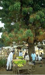
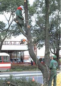
The colour plates give visual examples of different types of symptoms and damage associated with tree health problems. They are organized according to the classification of symptoms described in Table 3, Chapter 5. The captions describe the problem and give the name of the pest or other factors responsible for the symptoms. The host name and site are included in the caption. Colour plates can be searched by tree host and health problem in Annex 3.
Altered growth or development
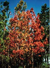
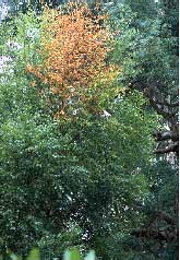
Plates 1A, 1B: COLOUR CHANGES IN CROWN
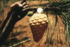
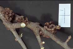
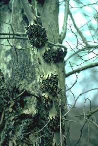
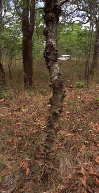
Plate 2: GALLS, SWELLINGS AND KNOTS
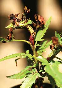
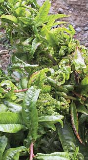
Plate 3: DISTORTION OF LEAVES AND STEMS
Plate 4: GROWTH STIMULATION
Plate 5: STUNTED OR REDUCED GROWTH
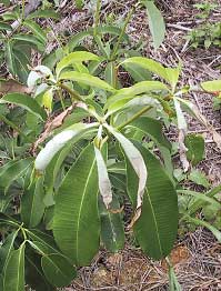
Plate 6: PREMATURE LOSS OF LEAVES OR STEMS
General death
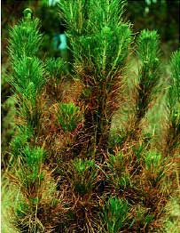
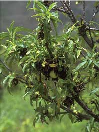
Plate 7: BLIGHT
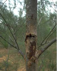
Plate 8: DIEBACK
Plate 9: WILT AND COLLAPSE
Localized death
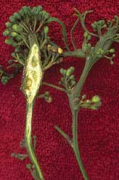
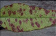
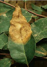
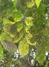
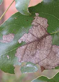
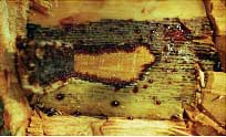
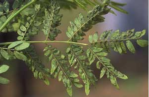
Plate 10: SPOTS AND LESIONS
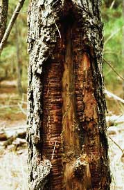
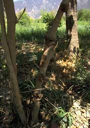
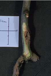
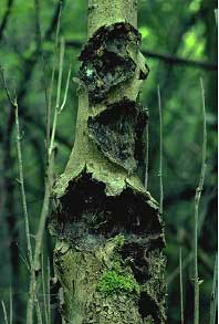
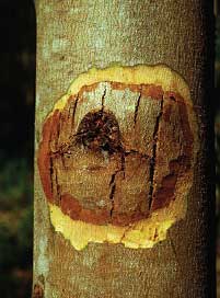
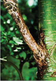
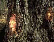
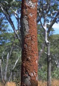
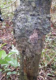
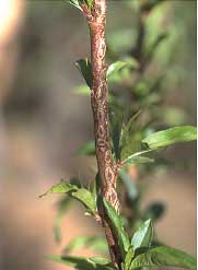
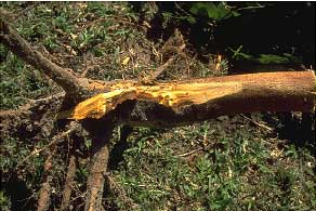
Plates 11A, 11B: CANKERS
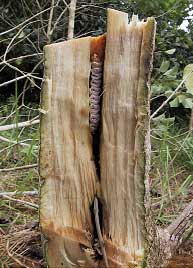
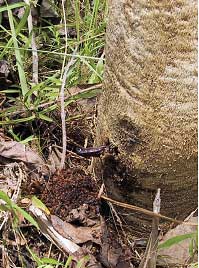
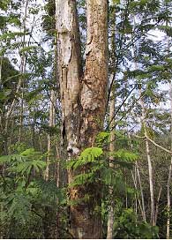
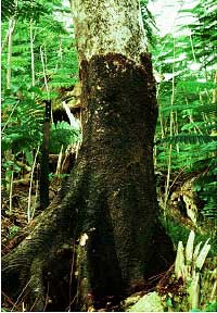
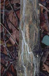
Plate 12: ROTS AND DECAYS
Physical evidence
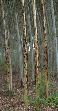
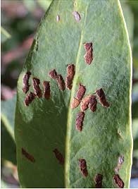
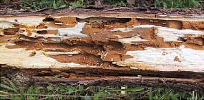
Plates 13A, 13B: DAMAGE BY INSECT AND ANIMAL FEEDING
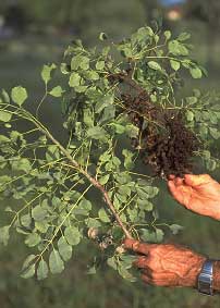
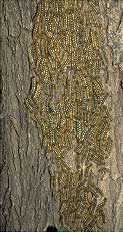
Plates 14A, 14B: PEST INFESTATION
Plates 15A, 15B: GENERAL DAMAGE
Plate 16: OTHER GROWTHS ON TREES
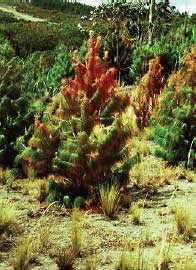
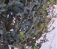
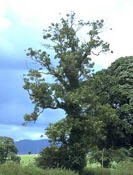
PLATE 1A
|
|||||
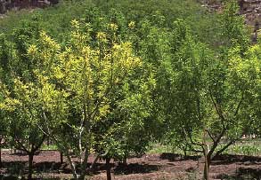
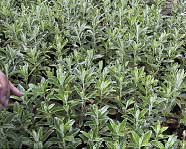
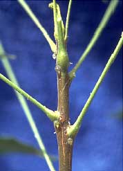
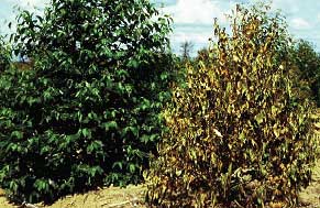
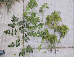
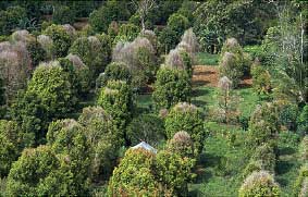
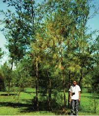
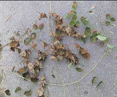
The examples in Plates 1A and 1B illustrate a wide variety of causes. Nutrient disorders and site factors are often said to be the cause of poor health, as determined by the appearance of crowns. But also look out for symptoms such as smaller leaves [1.2] and unusual patterns of leaf discolouration [1.5]. |
|||||
|
|
||||
|
|||||
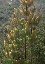
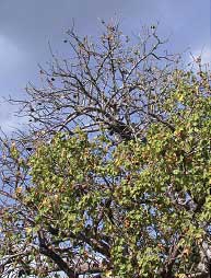
PLATE 1B |
||||
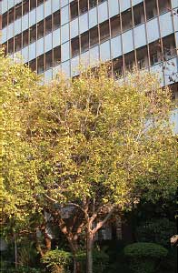
| Not all changes in colour indicate disease or insect attack [1.10]. Always compare the suspected problem with the condition or appearance of trees at different times of year. Nutrient imbalances in the soil are often blamed for colour changes and other altered growth responses, but little information exists about this for non-commercial tree species. |
||||
|
||||
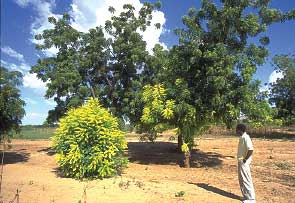
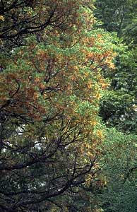
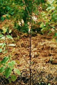
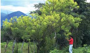
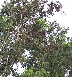
 1.9 Yellows disease (and dieback) due to phytoplasma disease. Melia azedarach, Santa Cruz de la Sierra, Bolivia. |
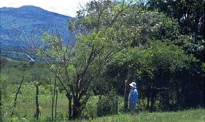
 1.10 Normal loss of foliage and colour change as the dry season begins. Toona ciliata, Zomba, Malawi. |
|||
 |
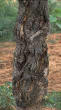
1.11 [left] Note the discolouration throughout the tree, the result of fungal canker damage caused by Cryphonectria cubensison the main stem (not visible in this photo). Eucalyptus sp., Brazil. | |||
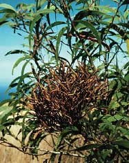
PLATE 2 |
|||||
The examples in Plates 1A and 1B illustrate a wide variety
of causes. Nutrient disorders and site factors are often said to be
the cause of poor health, as determined by the appearance of crowns.
But also look out for symptoms such as smaller leaves [1.2] and unusual
patterns of leaf discolouration [1.5]. |
|||||
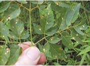
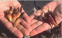
 2.1 Mite-induced galls causing significant damage in nurseries. Vangueria infausta, Gaborone, Botswana. |
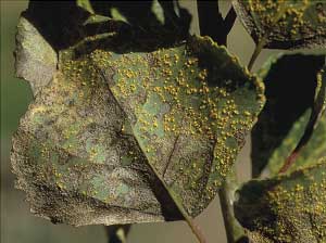
 2.2 Galls of the rust fungus, Atelocauda digitata. Acacia mangium, Kalimantan, Indonesia. |
||||
|
|||||
|
|||||
PLATE 3 |
|||||
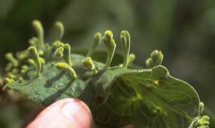
| A variety of pests can distort leaves and sometimes
young, still fleshy stems. The effects can be transient [3.2], as in
some incidences of insect feeding, but other altered growth forms can
be accompanied by serious losses [3.3]. |
|||||
 3.1 Virus disease produces distorted (shoestring) leaves on youngest stems. Gliricidia sepium, Vado Hondo, Guatemala. |
 3.2 Leaf curling and death of stems of unknown origin. Possibly due to insect feeding or virus disease. Uapaca kirkiana, Zomba, Malawi. |
||||
|
|||||
|
3.6 [left] Peach leaf curl, a highly distinctive fungus disease
caused by Taphrina deformans. Prunus persica, Epizana, Bolivia. |
||||
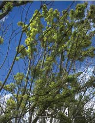
PLATE 4 |
|||||
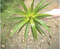
| Infection or pest attack can cause extra
or additional growth which is often weaker than normal and may lead to
dieback. |
|||||
|
|||||
|
|||||
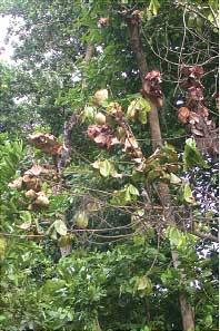
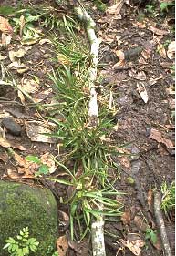
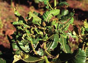
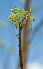
 4.6 Additional shoots formed with apparently smaller leaves. This type of symptom is difficult to interpret on unfamiliar trees. Suspected phytoplasma disease. Azanza garckeana, Gaborone, Botswana. |
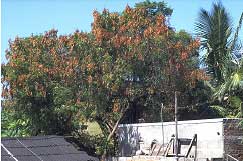
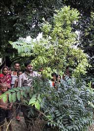 4.7 Spectacular growth at top of this tree is due to phytoplasma infection. Aphanamixis polystachya (Meliaceae), Dinajpur, Bangladesh. | ||||
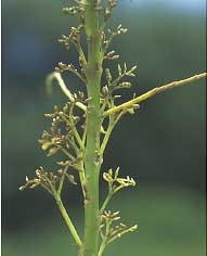
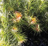
PLATE 5 |
||
Reduced growth may indicate poor growing conditions [5.1]. Major losses can occur when pests are not involved [5.4]. |
||
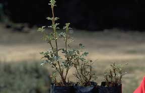
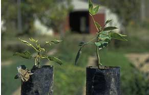 5.1 Compare reduced growth on left with seedling on right. Poor nutrition or lack of water are possible causes. No obvious indication of virus disease. Erythrina falcata, Cochabamba, Bolivia. |
 5.2 This native species grows up to 4,000 masl and beyond, but here it is performing poorly at 2,500 masl. Cause of problem is not known but could simply be poor nutrition. Polylepis incana, Cochabamba, Bolivia. |
|
 5.3 Gliricidia little leaf disease (phytoplasma). Internode length is greatly reduced giving this densely foliated stem. Gliricidia sepium, Zamorano, Honduras. |
 5.4 Greatly reduced internodes, barren lower stems and bunches of small leaves at the apex are known as giraffes' necks, a symptom of neem decline. Thought to be associated with drought. Azadirachta indica, near Sokoto, Nigeria. |
|
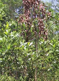
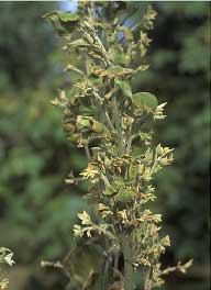
 5.5 Melia yellows disease. Stimulation of growth from main trunk. Leaves are much smaller and internode length is reduced, hence the compact bunches. These leaves soon die and fall away. Melia azedarach, Cochabamba, Bolivia. |
 5.6. Healthy foliage shown on the left and that caused by phytoplasma disease shown on the right. Melia azedarach, Bolivia |
|
PLATE 6 |
||
| Early drop of leaves is linked to pest [6.2] and non-pest [6.1] causes although in the former case trees would usually exhibit other symptoms such as root decay. The effect of abiotic influences such as drought is often temporary with trees becoming healthy as more rain falls. Pathogens and diseases have more fundamental and longer-term impacts on tree health. |
||
 6.1 Sparse crowns and early leaf drop as well as giraffe neck symptoms indicate neem decline. Thought to be associated with drought. Azadirachta indica, Damaturu, Nigeria. |
 6.2 Loss of leaves is due to phytophthora root disease. Alnus glutinosa, Sussex, UK. |
|
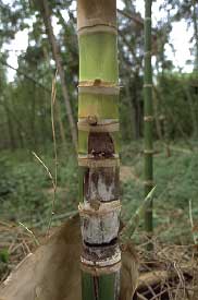
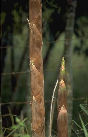 6.3 Premature death of culm sheaths is the first sign that culm tissues below are dying. Guadua angustifolia, Quindío, Colombia. |
 6.4 Loss of leaves is due to a bacterial wilt disease caused by Pseudomonas syzygii, known as Sumatra disease. Syzygium aromaticum, North Sumatra, Indonesia. |
|
 6.5 Loss of foliage (and yellowing) due to bacterial wilt caused by Ralstonia solanacearum. Casuarina equisetifolia, China. |
||
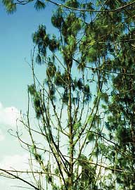
PLATE 7 |
|||||
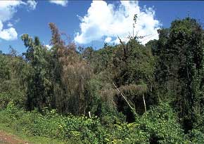
| Blight is a general term used to describe death of foliage or stems, often in the absence of any distinctive symptom. |
|||||
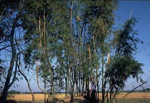 7.1 Bamboo blight, suspected fungal disease. Note brown, dead portions of blighted culms. Bambusa vulgaris, Chittagong, Bangladesh. |
 7.2 Dying bamboo in foreground which looks like blight, is due to flowering of bamboo. Arundinaria alpina, Ethiopia. |
||||
|
|||||
|
|||||
PLATE 8 |
|||||
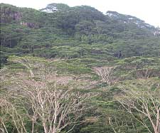
| Note that not all stems without leaves in the crown are necessarily dead. Dieback is associated with many different causes, from root and trunk diseases to the broader effects of poor growing conditions and waterlogging. |
|||||
 8.1 Note minor dieback at left due to little leaf disease (phytoplasma). The yellow foliage is also abnormal. Gliricidia sepium. Jutiapa, Guatemala. |
 8.2 Same tree as 8.1 photographed exactly one year later. Note more extensive dieback. |
||||
 8.3 Dieback due to unknown cause in street tree. Catalpa bignonioides, Baden-Baden, Germany. |
 8.4 Apparent dieback. Birds have stripped the foliage to make nests but stems may still be alive. Azadirachta indica, Garoua, Cameroon. |
||||
|
|||||
PLATE 9 |
|||||
| Wilting of foliage is often less distinctive in woody hosts when compared to the wilting of fleshy leaves of annual crops. Wilts are the result of internal blockages and are often accompanied by internal staining of stems [9.5]. Note that obtaining clear photos of wilt symptoms in crowns is often difficult; affected branches should be removed and photographed separately. |
|||||
 9.1 Wilt symptoms thought to be associated with fusarium root disease. Dalbergia sissoo, near Borga, Bangladesh. |
 9.2 Close-up of the wilt and "blighted" leaves shown in 9.1. |
||||
 9.3 Feeding by the tea mosquito, Helopeltis antonii, induces wilt-like symptoms. Azadirachta indica, India. |
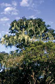
 9.4 Lethal yellowing disease of palms. Cocos nucifera, Florida, USA. |
||||
|
|||||
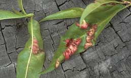
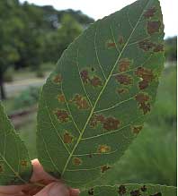
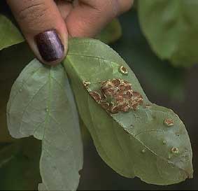
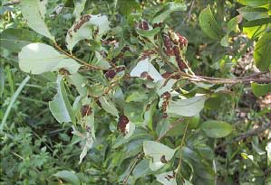
PLATE 10 |
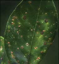
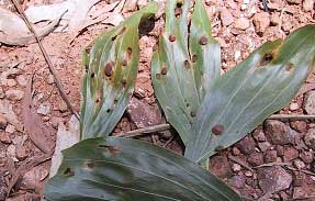
|||
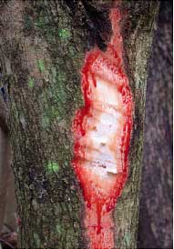
| Spots and lesions are common and it is not always easy to distinguish between insect feeding, fungi, bacteria, viruses and the effects of nutrient disorders. With stem lesions, carefully remove tissues to look for necrosis below [10.6]. |
|||
|
|||
|
|||
|
PLATE 11A |
|||
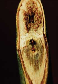
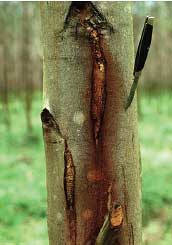
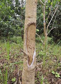
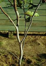
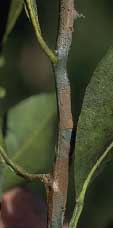
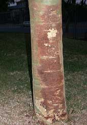
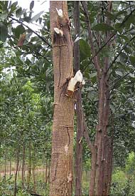
| Cankers can vary considerably in appearance: some have raised edges and cavities while others look more like galls or knots. The underlying tissues should be examined to see if there is necrosis and evidence of disease. |
|||
|
|||
|
PLATE 11B |
||||
| Some cankers do not have large cavities [11.7]. Note the features of gummosis on eucalypts [11.12 and 11.13]. |
||||
|
||||
|
||||
 |
11.13 Dark areas above exposed to show exudation of
liquid known as kino. |
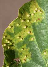
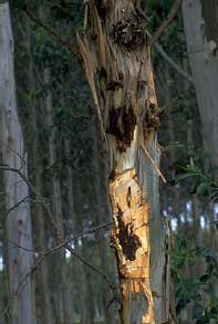
|||
PLATE 12 |
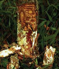
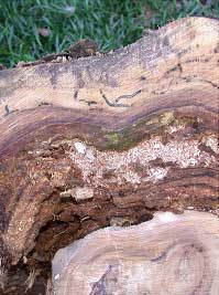
||||
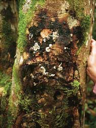
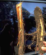
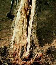
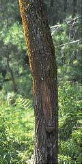
| Rots and decays are classified according to the place where they occur, e.g. heartrot [12.2] or according to colour [12.3] |
||||
 12.1 Suspected pythium root infection. Eucalyptus sp., KwaZulu, South Africa. |
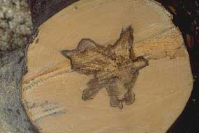 12.2 Suspected Ganodermasp. infection. Acer pseudoplatanus, France. |
|||
|
||||
|
||||
PLATE 13A |
||||
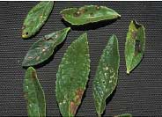
| Observing insects feeding on foliage is not always possible although some types of damage are unmistakable [3.7]. |
||||
 |
13.1 Psyllid infestation (Heteropsylla
cubana). Leucaena leucocephala, Nepal. |
|||
|
||||
|
||||
PLATE 13B |
|||
| Wilting of foliage in [13.11] and [13.13] is due not to systemic infection by pathogens but to animals feeding on the bark. Trees usually recover the following year. |
|||
|
|||
|
PLATE 14 |
|||
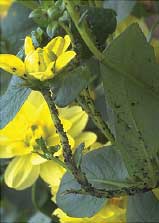
| Infestations are easy to observe and often cause concern, but their significance may be less than assumed. |
|||
|
|||
|
|||
|
PLATE 14B |
||||
| Observing insects feeding on foliage is not always possible although some types of damage are unmistakable [3.7]. |
||||
 14.10 Rust fungus (Melampsoraallii-populina). Populus sp., Cochabamba, Bolivia. |
 14.11 Rust fungus (Chaconia ingae) with raised pustules. Inga cylindrica, Santa Cruz, Bolivia. |
|||
 14.12 Gall mite (Aceria litchii) on lychee seen as "fur" on underside of leaves. Litchi chinensis, Thai Nguyen, Viet Nam. |
 14.13 Parasitic plant in crown as shown by the red flowers of Ligaria cuneifolia. Schinus molle, Santa Cruz, Bolivia. |
|||
|
||||
PLATE 15A |
|||
|
Unusual growth events [15.2] or damage [15.3] are best resolved by first looking for evidence of necrosis and then making enquiries about possible sources of mechanical damage. |
|||
|
|||
|
|||
|
PLATE 15B |
|||
| Bad pruning is a common source of future problems as the cut surfaces allow the entry of potential pathogens. Poor planting stock weakens future growth and makes trees more susceptible to insect attack. |
|||
|
|||
|
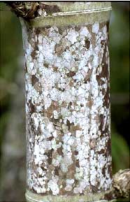
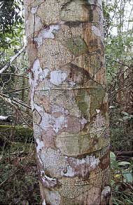
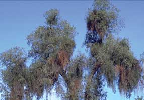
PLATE 16 |
|||
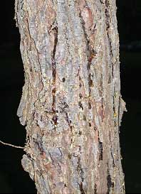
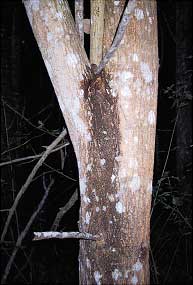
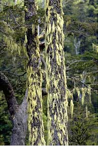
| Although plants and lichens do not cause direct harm to trees, they may have some effect or importance. Lichen patterns on stems may superficially resemble possible pathogenic fungi [16.6] |
|||
|
|||
|