


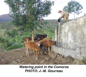
In February 2003, EMPRES was alerted by the National Veterinary Authorities of the Comoros that frequent mortalities had been recorded in the cattle population. A preliminary epidemiological investigation carried out in the field identified cattle imported from the United Republic of Tanzania as the possible source of infection.These animals had been introduced to the Comoros without previous sanitary control (quarantine).
More than 500 cattle died or were destroyed and 6 000 were at risk.
 |
 |
The salient clinical and pathological signs reported to EMPRES were as given in Table5.
TABLE 5 Clinical and pathological signs reported to EMPRES |
|
Clinical signs |
Pathological findings |
Peracute Acute Subacute |
Generalized hepatic necrosis in some cases |
The epidemiology and clinical signs observed in sick animals oriented suspicion towards a group of vector-borne diseases and more specifically towards bovine theileriosis which is endemic in East Africa. However, the samples collected could not confirm the presence of the disease |
In light of this emergency situation, the FAO Representation in Madagascar responded by initiating a letter of agreement with the Association comorienne des techniciens et infirmiers vétérinaires (ACTIV), a non-governmental organization, in order to provide urgent funds necessary to carry out the first preliminary field investigation. From the EMPRES side, Dr Jean-Marie Gourreau (Ecole nationale vétérinaire, Maisons-Alfort, France) was recruited to assist in the epidemiological investigation and went to the Comoros from 16 to 22 June 2003. During his mission, Dr Gourreau, in close collaboration with the national authorities and ACTIV, assessed the situation and highlighted the difficulty in establishing a definitive diagnosis. Little cooperation was obtained from livestock owners affected by the disease and only a few cases were available for clinical or pathological evaluation. The epidemiology and clinical signs observed in sick animals oriented suspicion towards a group of vector-borne diseases, and more specifically towards bovine theileriosis, which is endemic in East Africa. However, the samples collected could not confirm the presence of the disease.
Other aetiological considerations in the differential diagnosis included acute trypanosomiasis (Trypanosoma vivax), cowdriosis, babesiosis and rinderpest disease.
Without a final diagnosis to define an appropriate control strategy, additional samples were needed as the problem affecting cattle had subsided but not disappeared. A second mission was consequently organized. Vincent Martin, FAO-EMPRES Animal Health Officer (Infectious Disease Emergencies) travelled to the Comoros in November 2003 in collaboration with Dr Cheryl French, Veterinary Epidemiologist and APHIS representative for Africa. The principal objective of the mission was to identify precisely the causative agent responsible for the mortality in cattle.
The field mission visited newly affected villages in the northeast of the island (Moidja, Ngnadomboni and Mbeni). Twenty-one animals were clinically examined and three necropsies were performed. The following samples were also collected and sent to the Onderstepoort Veterinary Institute in South Africa:
 |
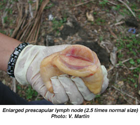 |
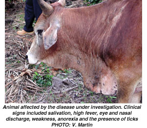
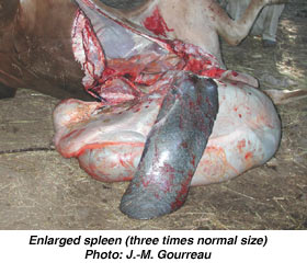
During the field investigation, it was observed that general pasture and feeding conditions were often very poor and not adequate to keep the animals in a good general condition. Sick animals had high temperatures (40-41.5°C), anorexia, weakness, ocular and nasal discharge, ptyalism, diarrhoea and enlargement of the prescapular lymph nodes. The cattle examined also showed heavy tick infestation. On post-mortem, the following signs were observed: splenic enlargement, enlarged prescapular lymph nodes, sometimes showing oedematous and haemorrhagic lesions, dry fore stomach omasum and reticulum, and pasty ruminant contents. In one young steer petechial haemorrhages were seen on the kidney.
The first results from the Ondestepoort Veterinary Institute were suggestive of theileriosis, in association with other diseases such as babesiosis. The plants collected were not found to be toxic. More details will be provided in the next issue of the Bulletin.
