![]()
![]()
Avian influenza (AI) has been recognized as a highly lethal generalized viral disease of poultry since 1901. In 1955, a specific type (A) of influenza virus was identified as the causal agent of what was then called “fowl plague”. It has since been found that AI viruses cause a wide range of disease syndromes, ranging from severe to mild, in domestic poultry.
The influenza viruses that constitute the family Orthomyxoviridae are classified into types A, B or C based on differences between their nucleoprotein and matrix protein antigens. AI viruses belong to type A. Influenza viruses are further categorized into subtypes according to the antigens of the haemagglutinin (H) and neuraminidase (N) projections on their surfaces. There are 14 haemagglutinin subtypes and 9 neuraminidase subtypes of influenza A viruses, and AI viruses have representatives in all of these subtypes. However, to date all highly pathogenic AI viruses that cause generalized rather than respiratory disease belong to either the H5 or H7 subtype. The pathogenicity of AI viruses is correlated to the ability of trypsin to cleave the haemagglutinin molecule into two subunits. Highly pathogenic strains of H5 and H7 viruses have several amino acid residues at the cleavage site. Trypsin sensitivity and amino acid sequencing can be used diagnostically to determine whether or not an isolated virus is potentially pathogenic.
Influenza A viruses infecting poultry can be divided into two distinct groups on the basis of their ability to cause disease in chickens. The most virulent viruses cause “fowl plague”, now termed “highly pathogenic avian influenza” (HPAI), in which mortality may be as high as 100 percent. These viruses have been restricted to subtypes H5 and H7, although not all viruses of these subtypes cause HPAI. Other viruses cause a much milder, primarily respiratory, disease designated “low pathogenic avian influenza” (LPAI), which nevertheless may be exacerbated by other infections or environmental conditions, resulting in a much more serious disease.
|
H. WAGNER |
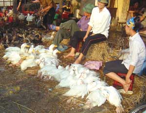
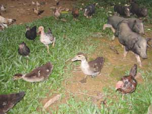
Domestic fowl, ducks, geese, turkeys, guinea fowl, quail and pheasants are susceptible. Disease outbreaks occur most frequently in domestic fowl and turkeys. A particular strain may produce severe disease in turkeys, but not in chickens or any other avian species. Therefore, it would be impossible to generalize on the host range for HPAI, for it may vary with the isolate. This assumption is supported by reports of farm outbreaks where only a single avian species of several species present on the farm has become infected. Many species of wild birds, particularly waterfowl and sea birds, are also susceptible, but infections in these birds are generally subclinical.
The immediate source of infection for domestic poultry can seldom be ascertained, but most outbreaks probably start with direct or indirect contact of domestic poultry with waterbirds. Many of the strains that circulate in wild birds are either non-pathogenic or mildly pathogenic for poultry. However, a virulent strain may emerge either by genetic mutation or by reassortment of less virulent strains. Scientific evidence indicates that the former mechanism occurred in 1983-87 in the eastern United States of America. Swine appear to be important in the epidemiology of infection of turkeys with swine influenza virus when they are in close proximity.
|
R. WEBB |
Other mammals do not appear to be involved in the epidemiology of HPAI. However, during the 2004 HPAI crisis in Southeast Asia, infections of cats were reported. The infection of humans with an H5 avian influenza virus in China’s Hong Kong Special Administrative Region in 1997 has resulted in a reconsideration of the role of the avian species in the epidemiology of human influenza.
Once AI is established in domestic poultry, it is a highly contagious disease, and wild birds are no longer an essential ingredient for spread. Infected birds excrete the virus in high concentration in their faeces and also in nasal and ocular discharges. Once introduced into a flock, the virus is spread from flock to flock by the usual methods involving the movement of infected birds, contaminated equipment, egg cartons, feed trucks and service crews, to mention a few. The disease generally spreads rapidly in a flock by direct contact, but on occasion spread is erratic.
Airborne transmission may occur if birds are in close proximity and with appropriate air movement. Birds are readily infected via instillation of the virus into the conjunctival sac, nares or trachea, and the virus is transmitted among waterfowl by cloacal drinking. Preliminary field and laboratory evidence indicates that the virus can be recovered from the yolk and albumen of eggs laid by hens at the height of the disease. The possibility of vertical transmission is unresolved; however, it is unlikely infected embryos could survive and hatch. Attempts to hatch eggs in disease isolation cabinets from a broiler breeder flock at the height of disease have failed to result in any AI-infected chickens. This outcome does not mean that broken contaminated eggs could not be the source of virus to infect chicks after they hatch in the same incubator. The hatching of eggs from a diseased flock would likely be associated with considerable risk.
|
D. SWAYNE, UNITED STATES |
The incubation period is usually three to seven days, depending upon the strain, the dose of inoculum, the species and the age of the bird.
The clinical signs are very variable and are influenced by factors such as the virulence of the infecting virus, species affected, age, sex, concurrent diseases and environment.
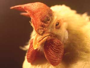
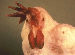
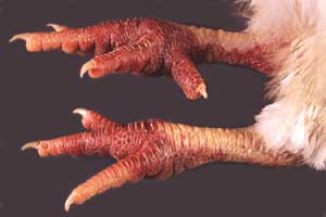
In virulent (or highly pathogenic) AI, the disease appears suddenly in a flock, and many birds die either without premonitory signs or with minimal signs of depression, inappetence, ruffled feathers and fever. Other birds show weakness and a staggering gait. Hens may at first lay soft-shelled eggs, but soon stop laying. Sick birds often sit or stand in a semi-comatose state with their heads touching the ground. Combs and wattles are cyanotic and oedematous and may have petechial or ecchymotic haemorrhages at their tips. Profuse watery diarrhoea is frequently present, and birds are excessively thirsty. Respiration may be laboured. Haemorrhages may occur on unfeathered areas of skin. The mortality rate varies from 50 to 100 percent. In broilers, the signs of disease are frequently less obvious with severe depression, inappetence and a marked increase in mortality being the first abnormalities observed. Oedema of the face and neck and neurological signs such as torticollis and ataxia may also be seen. The disease in turkeys is similar to that seen in layers, but it lasts two or three days longer and is occasionally accompanied by swollen sinuses. In domestic ducks and geese the signs of depression, inappetence and diarrhoea are similar to those in layers, though frequently with swollen sinuses. Younger birds may exhibit neurological signs. Ducks infected with HPAI and excreting the virus may not show any clinical signs or lesions.
|
D. SWAYNE, USDA |
Birds that die of peracute disease may show minimal gross lesions, consisting of dehydration and congestion of viscera and muscles. In birds that die after a prolonged clinical course, petechial and ecchymotic haemorrhages occur throughout the body, particularly in the larynx, trachea, proventriculus and epicardial fat, and on serosal surfaces adjacent to the sternum.
There is extensive subcutaneous oedema, particularly around the head and hocks. The carcass may be dehydrated. Yellow or grey necrotic foci may be present in the spleen, liver, kidneys and lungs. The air sac may contain an exudate. The spleen may be enlarged and haemorrhagic.
AI is characterized histologically by vascular disturbances leading to oedema, haemorrhages and perivascular cuffing, especially in the myocardium, spleen, lungs, brain and wattles. Necrotic foci are present in the lungs, liver and kidneys. Gliosis, vascular proliferation and neuronal degeneration may be present in the brain.
|
D. SWAYNE, USDA |
The following diseases must be considered in the differential diagnosis of HPAI:
Other diseases causing sudden high mortality:
Newcastle disease
infectious laryngotracheitis
duck plague
acute poisonings
Other diseases causing swelling of the combs and wattles:
acute fowl cholera and other septicaemic diseases
bacterial cellulitis of the comb and wattles
Less severe forms of the disease may be confused with, or complicated by, many other diseases with respiratory or enteric signs. AI should be suspected in any disease outbreak in poultry that persists despite the application of preventive and therapeutic measures for other diseases.
Specimens required
|
R. WEBB |
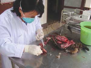
Specimens should be collected from at least six birds. Preferably, three should be birds showing signs of the acute disease, and the other three may be recently dead. Swabs of tracheal and cloacal contents, brain and heart blood should be collected aseptically for confirmational and differential diagnostic purposes. The material collected on the swabs should be mixed into 3 ml aliquots of transport medium in sterile bottles and the swabs discarded. The transport medium may be sterile brain-heart infusion broth containing 5 000 units of penicillin and 5 000 µg of streptomycin per millilitre, or equal parts of glycerol and phosphate-buffered saline with the same antibiotics added. Tracheal and cloacal swabs should also be collected from selected live birds in the flock. At autopsy, unpreserved specimens of brain, trachea, spleen and intestinal contents should be collected for isolation of the virus. Impression smears should be made of internal organs, including kidney and pancreas, for detection of viral antigen by immunofluorescent techniques.
Blood samples should be collected for serum. Samples should be taken from several birds in the flock.
Transport of specimens
Unpreserved tissues and swab material should be chilled and forwarded on water ice or with frozen gel packs. If delays of more than 48 hours are expected in transit, these specimens should be frozen and forwarded with dry ice.
Laboratory procedures
|
S. DESVAUX |
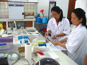
AI virus is most commonly isolated by inoculation of swab material or tissue homogenates into nine- to eleven-day-old embryonated chicken eggs by the allantoic sac route. The embryos may or may not die, but in any case the presence of the virus can be detected by haemagglutinin tests on harvested allantoic fluid. Its identity is confirmed by agar gel diffusion or haemagglutination inhibition tests using specific antiserum. Rapid diagnosis can be made by the detection of viral antigen in tissue impression smears using immunofluorescence, or by antigen detection enzyme-linked immunosorbent assay (ELISA) on tissue homogenates. The pancreas and kidneys are the organs in which antigen is most often demonstrable. Isolates of the virus can be serotyped to determine their haemagglutinin and neuraminidase subtypes. Pathogenicity tests are carried out by inoculating four- to six-week-old chickens intravenously or into the caudal thoracic air sac with an inoculum prepared from infectious allantoic sac fluid. A pathogenicity index is determined from the number of healthy, sick, paralysed and dead birds observed each day for ten days post-inoculation. In vitro tests, based on the ability of the virus to produce plaques in cell cultures in the absence of trypsin, are also useful for pathotyping strains of the virus. However, polymerase chain reaction and gene sequencing procedures can be used for rapid determination of the pathogenic potential of an AI virus. This is an important aspect in determining the role of the virus in the disease seen in the field. Group-specific antibody can be detected by ELISA in serum samples from birds two weeks or more after they first show clinical signs.
Once the subtype of the virus has been determined, haemagglutination inhibition tests can be used for active surveillance to detect antibodies.
Alexander, D.J. 2000. A review of avian influenza in different bird species. Vet. Microbiol., 74(1-2): 3-13.
Geering, W.A., Forman, A.J. & Nunn, M.J. 1995. Exotic diseases of animals, a field guide for Australian veterinarians. Canberra, Australian Government Publishing Service.
World Organisation for Animal Health (OIE) Web site
![]()
![]()