![]()
![]()
![]()
Clinical findings
Discussion
Bibliography
Short communications/brèves communications/ communicaciones breves
R.O. Ramadan, N.H. Hashim and A.A.E. Bukhari
Dr Ramadan is an Associate Professor of Surgery, Department of Clinical Studies, College of Veterinary Medicine and Animal Resources. King Faisal University, Box 1757, Al Ahsa; Dr Hashim is an animal health expert, Veterinary Training Centre, Box 573, Al Ahsa; Dr Bukhari is a member of the Department of Clinical Studies, College of Veterinary Medicine and Animal Resources as well as Director of the Veterinary Teaching Hospital and the Agriculture and Veterinary Training and Research Station, King Faisal University, Al Ahsa, Saudi Arabia
Four cases of carpal hygroma in sheep in the Kingdom of Saudi Arabia with Brucella antibody titres ranging from 160 immunizing units (IU) to 640 IU were clinically, radiographically and bacteriologically investigated. Brucella melitensis was isolated from one animal and swellings of the carpal regions of the other three animals under investigation were surgically removed. Carpal hygroma is a false bursa situated over the anterior aspect of the carpal joint. The condition was reported in cattle (Jensen and Mackey, 1965) and in horses (Veenendaal, Spiers and Harrison, 1981). In cattle it was described as localized lesion related to Brucella infection (Balbo, Nobil and Guercio, 1969; Shigidi and Razig, 1973) and the presence of hygromas was considered evidence of brucellosis in the herd.
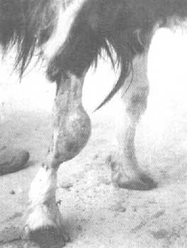
Examination of four sheep (three rams and one ewe) afflicted with unilateral carpal hygroma revealed a fluctuating swelling over the anterior part of the right carpus in three cases and over the left carpus in the fourth. All animals were from flocks with a history of abortion and placenta retention. The ewe had a normal delivery and had not previously shown any problems. Body temperature was normal in all cases. Serum and hygroma fluid samples from the four cases were aseptically collected for serological and bacteriological investigations.
Radiographic examination
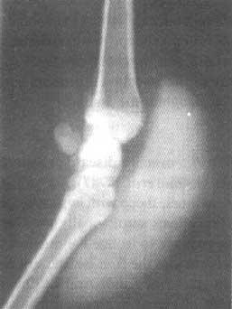
Plain and contrast radiographs of the affected carpi were taken in a routine manner. For contrast radiographs, 5 to 10 ml of 70 percent w/v solution of sodium iothalamate were aseptically injected into the swellings. The fluid was dispersed and then mediolateral radiographs were taken. The contrast agent remained in the soft tissue swelling which did not communicate with the carpal sheath or the joint capsule and was separated from the carpal bones by a clear space (see Figs 1 and 2).
Serological and bacteriological examinations
The serum samples from the four animals were examined using the slide agglutination test with Rose Bengal stained antigen and the tube agglutination test with Brucella abortus polyvalent antigen. Each of the hygroma fluid samples were cultured in two Brucella agar plates and incubated at 37°C in 10 percent carbon dioxide atmosphere and air for four days. Brucella-like colonies were subjected to serological and biochemical tests as shown in Table 1.
1. Tests carried out on Brucella-like colonies
Tests effectués sur des colonies de germes du genre Brucella
Prueba realizada en colonias del tipo de las de Brucella
|
CO2 requirement |
H2S production |
Growth in presence of: |
Agglutination in polyvalent antiserum | |
|
|
|
Basic fuchsine 1/25 000 |
Thionine 1/50 000 |
|
|
- |
- |
+ |
+ |
+ |
2. Titres of sheep under examination
Titres d'anticorps chez les moutons examinés
Examen de títulos de ovejas
|
Number |
Breed |
Sex |
Serum antibody titre |
|
1 |
Najdi |
Male |
160 IU |
|
2 |
Najdi |
Male |
320 IU |
|
3 |
Awassi |
Male |
160 IU |
|
4 |
Najdi |
Female |
640 IU |
1. Carpal hygroma in a sheep - Mouton atteint d'un hygroma carpien - Oveja con higroma carpiano
Results and treatment
The four sheep were serologically positive for both Rose Bengal and the tube agglutination test with antibody titres ranging from -160 to 320 IU in serum samples of the rams and 640 in the ewe's serum (see Table 2). Hygroma fluid samples from the three rams showed no growth on culture. The sample from the ewe showed typical Brucella-like colonies in both 10 percent carbon dioxide atmosphere and air on the second day of incubation. From the results of the biochemical and serological tests (see Table 1), it was concluded that the organism was Brucella melitensis.
Surgery
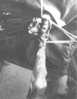
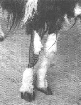
Surgical excision of the hygromas containing sterile fluid was performed in three animals. Each animal was sedated with Xylazine (2 percent), given intravenously in the dose of 0.2 mg/kg body weight. This was followed by local infiltration analgesia using lignocaine hydrochloride. The swellings were carefully dissected in each case. Skin sutures were routine (see Figs 3 and 4). The wound healed by first intention without any undue delay and the animals were discharged. The fourth sheep, with Brucella organisms :in its synovial fluid, was not operated on because of the possibility of the infection spreading and the owner was advised to slaughter the animal.
Brucellosis caused by B. melitensis is a disease prevalent in animals in Saudi Arabia (Hashim et al., 1987). According to the experience of these authors, sheep with hygroma are not uncommon among brucellosis serologically positive flocks but no attempts were made to isolate the organism from the site. This report provides the first isolation of B. melitensis from sheep hygroma in Saudi Arabia. Other aetiological considerations of carpal hygromas included repeated trauma or the infection of the carpal joint with other pathogenic micro-organisms.
In the present study the joint capsule was not involved in any of the four cases, and this gave a similar picture to equine hygromas (Veenendaal, Spiers and Harrison, 1981).
Treatment of hygromas included aspiration and injection of corticosteroid preparation, drainage procedure and insertion of seton or excision. Excision of hygromas has been performed on horses but requires general anaesthesia, which is often tedious and might produce extensive fibrosis. Contrast radiographs are essential to exclude joint involvement.
The positive sera and negative organisms in the absence of external lesions-might reflect immunity as a result of vaccination or foci elsewhere in the body. However, verification of the latter point would require slaughtering of the animals. Brucellosis is an endemic problem in this region but the incidence of hygromas among the reactors is yet to be recorded. The presence of hygromas in sheep is evidence of brucellosis in the flock as it is for cattle (Balbo, Nobit and Guercio, 1969). Therefore, animals suffering, from hygromas represent a potential hazard to both the flock and humans who come into contact with them.
Balbo, S.M, Nobil, I. & Guercio, V. 1969. Hygromas in cattle infected with Brucella abortus: their importance in the diagnosis of the disease. Vet. Ital., 20: 709-715 (cit. Vet. Bull., 40: 535).
Hashim, N.H., Galil, G.A., Hulaibi, M.A. & Al-Saleem, F.M. 1987. The incidence of brucellosis and species of Brucella organisms isolated from animals in Al Hassa. World Anim. Rev. (61)32-35
Jensen, R. & Mackey, D.R. 1965. Diseases of feedlot cattle. Philadelphia. Lea and Febiger. 91 pp.
Shigidi, M.A. & Razig, S.A. 1973. Isolation of Brucella abortus from a knee hygroma in a bull. S.J. Vet. Sci. & Anim. Hasb., 14: 33-35.
Veenendaal, J.C., Spiers, V.C. & Harrison, I. 1981. Treatment of hygromata in horses. Australian Vet. J.,57(11)513-514.
![]()
![]()
![]()