Part I: Detection and identification of specific grapevine diseases or pathogens
True
virus diseases
Rugose wood complex
Viroid diseases
Virus-like diseases
Diseases induced by phloem- and xylem-limited
prokaryotes
Grapevine degeneration - fanleaf
Grapevine degeneration - European nepoviruses
Grapevine decline - American nepoviruses
Leafroll
Grapevine degeneration - fanleaf
G.P. Martelli
CAUSAL AGENT
The causal agent of fanleaf is grapevine fanleaf nepovirus (GFLV), a mechanically transmissible virus with isometric particles 30 nm in diameter and a bipartite genome (Quacquarelli et al., 1976). GFLV populations are remarkably homogeneous serologically, so that natural serological variants are rarely found when screening is done by conventional serological techniques (Saving, Chérif and Martelli, 1985). Serological differences are detected, however, by monoclonal antibodies (Huss et al., 1987). Biological variants differing according to the symptoms induced in artificially inoculated herbaceous hosts or in naturally infected grapevines are many.
GEOGRAPHICAL DISTRIBUTION
The disease and the causal virus have been recorded from all major grapevine-growing areas of the world. The level of infection is often very high, so that cultivars grown in certain viticultural districts are totally diseased.
ALTERNATE HOSTS
No alternate host is known. GFLV has never been found naturally in any wild or cultivated plant species other than Vitis vinifera and American Vitis species.
FIELD SYMPTOMS
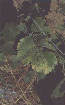
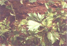
Fanleaf degeneration is characterized by two distinct syndromes deriving from a differential reaction of the hosts to biologically distinct strains of the causal virus. Vein banding, another syndrome traditionally thought to be caused by chromogenic GFLV strains, may have a different aetiology, perhaps viroidal, as discussed under Viroid diseases.
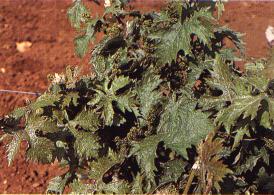
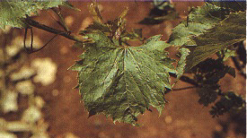
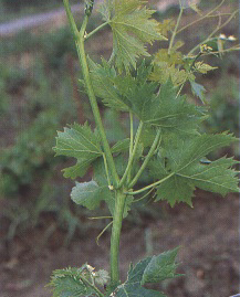
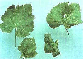
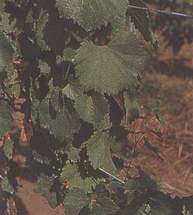
Infectious malformations (fanleat proper)
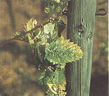
Distorting virus strains may cause vines to be
stunted or less vigorous than normal. Leaves are variously and
severely distorted, asymmetrical, cupped and puckered and exhibit
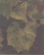
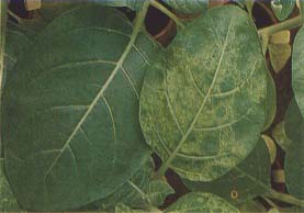
acute dentations (Figures 1 and 2). Chlorotic mottling may
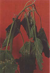
sometimes accompany foliar deformations (Figure 3). Canes are
also malformed, showing abnormal branching, double nodes,
different length or exceedingly short internodes, fasciations and
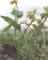
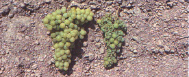
zigzag growth (Figures 4 and 5). Bunches are reduced in number
and size, ripen irregularly and have shot berries and poor berry
setting (Figure 6). Foliar symptoms develop early in the spring
and persist through the vegetative season, although some masking
may occur in summer.
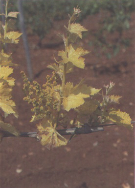
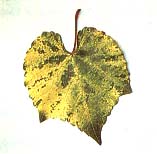
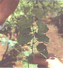
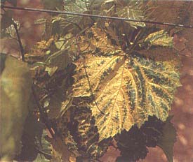
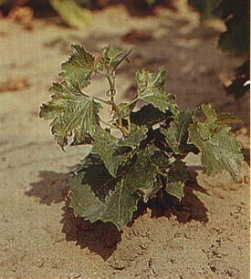
Yellow mosaic
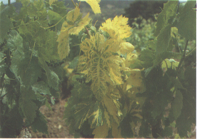
Yellow mosaic is caused by chromogenic virus
strains. Affected vines show chrome-yellow discolorations that
develop early in the spring and may affect all vegetative parts
of the vines (leaves, herbaceous shoot axes, tendrils and
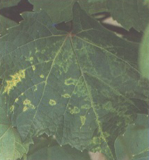
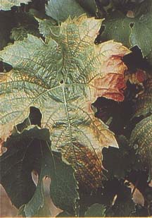
inflorescences) (Figure 7). Chromatic alterations of the leaves
vary from a few scattered yellow spots, sometimes appearing as
rings and lines, to variously extended mottling of the veinal
and/or interveinal areas, to total yellowing (Figures 8 to 11).
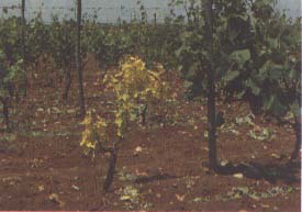
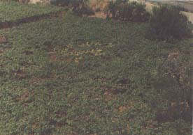
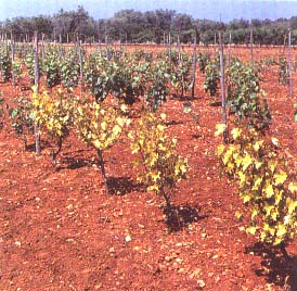
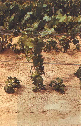
In spring, affected plants in a vineyard can readily be spotted
from a distance (Figure 12). Malformations of leaves and canes
are usually not prominent, but clusters may be smaller than
normal and may have shot berries. In hot climates the newly
produced summer vegetation has a normal green colour, while the
yellowing of the old growth turns whitish and tends to fade away
(Figure 13).
Symptoms induced by distorting virus strains show up equally well under field and greenhouse conditions, whereas chrome-yellow alterations evoked by chromogenic strains may not develop in greenhouses.
NATURAL SPREAD
Spread at a site (i.e. within a vineyard or between adjacent vineyards) is mediated by nematodes. Two longidorid nematode vectors of GFLV are known: Xiphinema index and Xiphinema italiae (Hewitt, Raski and Goheen, 1958; Cohn, Tanne and Nitzany, 1970). The former species is by far the more efficient vector under natural and experimental conditions. Although not all X. index populations are equally efficient in transmitting virus isolates (Catalano, Roca and Castellano, 1989), this nematode is to be regarded as the major, if not the only, natural and economically important GFLV vector. It has a limited range of alternate natural hosts (e.g. fig, mulberry, rose), but these hosts are immune to GFLV. No natural virus reservoirs are known other than grapevines. GFLV persists in volunteer plants and in the roots of lifted vines that remain viable in the soil, constituting an important source of inoculum. Transmission of GFLV through grapevine seeds has been reported (Lazar, Köber and Lehoczky, 1990), but it has negligible epidemiological significance.
Long-distance spread is passively but efficiently effected through dissemination of infected propagating material (budwood and rooted cuttings of rootstocks) (Martelli, 1978).
DETECTION
Field symptoms are often obvious enough to allow the detection of diseased vines. Difficulties may be encountered, however, even by keen and experienced observers, in the presence of tolerant varieties and/or infections by mild virus strains. Observations for symptoms should be carried out twice during the vegetating season: in midspring for growth abnormalities, deformation and chromatic alterations of the foliage; and at leaf shedding for abnormalities of the canes.
IDENTIFICATION
Indexing by graft transmission
Although none of the Vitis species used as indicators is immune to GFLV, the quickest and most typical responses are given by Vitis rupestris St George. In greenhouse indexing (chip-budding, green grafting) at 22 to 24°C shock symptoms appear as soon as three to four weeks after grafting. The symptoms consist of chlorotic spots, rings and lines (Figure 14), sometimes accompanied by malformations and localized necrosis of the tissues. These symptoms are transient. They are followed by chronic reactions that, for distorting virus strains, consist of reduced growth and severely deformed leaves with prominent teeth (Figure 15). Chronic symptoms develop within two to three months from inoculation and persist throughout the vegetating season (Hewitt et al., 1962; Martelli and Hewitt, 1 963).
Field indexing (cleft or whip grafting) reactions are slower. Symptoms appear during the first year of vegetation. They are of the chronic type for distorting virus strains but consist of various patterns of yellow discolorations, sometimes accompanied by leaf deformity (Figure 16) for chromogenic virus strains. With certain virus sources yellowing may appear in the second year after inoculation. Yellow discolorations do not usually develop in greenhouse indexing.
Indicators other than V. rupestris may react to GFLV infections with chronic-type responses. These appear in the first or second year after grafting and consist of deformations and/or bright yellowing of the foliage. The type and severity of the symptoms vary with the nature and virulence of the virus strain.
Transmission to herbaceous hosts
GFLV is readily transmitted to herbaceous hosts
by inoculation of expressed sap, provided that some precautions
are observed. The best inoculum sources are young, tender
symptomatic leaves of the spring flush collected directly in the
field or from greenhouse-grown cuttings. Young, succulent roots
are equally good, if not superior, sources of inoculum. Suitable
root material is readily obtained by forcing grape cuttings to
root in sand in a tray or bench heated to 24 to 25°C. Old leaves
collected in the field in summer or autumn. especially in hot
climates, constitute a poor source of virus, even if they show
symptoms. Their use may result in repeated failures to transmit
the virus.
For inoculation, 3 to 5 g of tissues (either leaves or roots) are ground in a chilled mortar together with an equal volume of one of the extraction media described in Part II. Media containing nicotine are recommended when the inoculum consists of leaves, especially if they are aged, whereas ordinary phosphate buffer is quite suitable for young root tips.
The herbaceous host range of GFLV is fairly wide, comprising some 50 species in seven dicotyledonous families (Dies, 1963; Hewitt et al., 1962; Martelli and Hewitt, 1963 ; Taylor and Hewitt, 1964). Differential diagnostic hosts are:
Whereas the above hosts are infected symptomatically by most virus isolates, many additional herbaceous indicators, such as Phaseolus vulgaris, Cucumis sativus and Petunia hybrida, are more selective in their susceptibility, being infectible by a smaller number of strains.
Presence of trabeculae (endocellular cordons)
Trabeculae are intraxylematic ribbon-like inclusions that
cross tracheary elements radially. These structures are readily
observed in crosssectioned basal internodes of green or mature
canes. Sections made by hand with a razorblade are mounted
without staining in a 1:1 mixture of water and glycerol and
observed at low power undera light microscope.
Freshlycross-sectioned canes can also be observed directly with a
good (10 to 12x) magnifying lens. Trabeculae are more frequently
found in American rootstocks than in European scion varieties.
Their presence can be taken as evidence of viral infection, but
their absence does not guarantee freedom from GFLV.
Serology
Immunodiffusion in agar gel is not applicable to leaf extracts of GFLV-infected grapevines, regardless of the severity of symptoms shown and the conditions under which plants are grown (field or greenhouse). Positive reactions are more readily and consistently obtained using crude sap expressed directly, without addition of buffers, from leaves of systemically invaded herbaceous hosts. Young upper leaves with strong symptoms are the best source of antigen.
ELISA. Immunoenzymatic procedures (see details in Part III) are now routinely employed for detection of GFLV in field- or greenhousegrown vines (Bovey, Brugger and Gugerli, 1980; Engelbrecht, 1980; Walteretal., 1984).Sources of antigens can be buds, roots, leaves and wood shavings, the last two being the most commonly used. Reactants can be either polyclonal antibodies or virus-specific or strain-specific monoclonalantibodies(Hussetal.,1986,1987).
The amount of plant material needed can be as low as 100 to 200 ma. However, it is customary to use 1 g of leaf tissues and 0.5 g of wood shavings. These are obtained by removing the bark and scratching, with a scalpel or a razorblade, the cortex of mature canes freshly harvested or cold-stored at 4 to 6°C (Walter and Etienne, 1987). Tissue samples, regardless of whether taken from leaves or cortex, are ground in PBS-Tween containing 2 percent PVP and 1 to 2.5 percent nicotine. Addition of nicotine is considered of paramount importance if tests are to be made from leaf tissues. No nicotine is needed if the extraction medium is made up of 0.1 M Tris-HCI buffer, pH 8.2, containing 0.8 percent NaCI and 2 percent PVP (Walter and Etienne, 1987).
The advantage of wood shavings over leaves is twofold: they can be used throughout the year without the apparent loss of efficiency that arises because of seasonal variations of antigen titre in vegetating organs; and they give low and consistent background readings. If leaves are employed, it is important, if not mandatory, to use extracts of the same rootstock or European scion variety as negative and positive controls. There is, in fact, a marked difference in background readings between American rootstocks and European cultivars and, among V. vinifera varieties, between those with hairy and those with glabrous leaves.
Immune electron microscopy (IEM and lSEM). GFLV can be detected by ISEM following the procedure outlined in Part III. Consistent and satisfactory results are obtained in spring from upper symptomatic leaves (Bovey, Brugger and Gugerli, 1980; Russo, Martelli and Savino, 1980).
Molecular hybridization
Cloned cDNA probes have been prepared to genomic (RNA-1 and RNA-2) and satellite RNAs of GFLV. These probes were successfully used for identification of GFLV infections in field-grown grapevine leaf extracts denatured with formaldehyde (Fuchs et al., 1991) and processed as described in Part III.
SANITATION
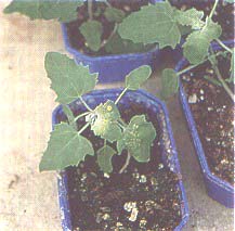
Sanitary selection and heat therapy together are powerful tools for reducing the incidence of fanleaf in newly established vineyards. Virusfree material (Figure 20) is readily obtained through conventional (Goheen and Luhn, 1973) or modified (Stellmach, 1980) heat therapy applications, micrografting and in vitro meristem tip or shoot-tip culture (Barlass et al., 1982).
REFERENCES
Barlass, M., Skene, K.G.M., Woodham, R.C. & Krake, R.C. 1982. Regeneration of virus-free grapevines using in vitro apical culture. Ann. Appl. Biol., 101: 291-295.
Bovey, R., Brugger, J.J. & Gugerli, P. 1980. Detection of fanleaf virus in grapevine tissue extracts by enzyme-linked immunosorbent assay (ELISA) and immune electron microscopy (IEM). Proc. 7th Meet. ICVG, Niagara Falls, NY, USA, 1980, p. 259-275.
Catalano, L., Roca, F. & Castellano, M.A.1989. Efficiency of transmission of an isolate of grapevine fanleaf virus (GFV) by three populations of Xipizinema index (Nematode: Dorylaimidae). Nematol. Mediterr., 17: 13- 15.
Cohn, E., Tanne, E. & Nitzany, F. 1970. Xiphinema italiae: a new vector of grape fanleaf virus. Phytopathology, 60: 181 - 182.
Dias, H.F. 1963. Host range and properties of grapevine fanleaf and grapevine yellow mosaic viruses. Ann. Appl. Bial., 5 I: 85-95.
Engelbrecht, D.J. 1980. Indexing grapevines for grapevine fanleaf virus by enzymelinked immunosorbent assay. Proc. 7th Meet. ICVG, Niagara Falls, NY, USA, 1980, p. 277282.
Fuchs, M., Pinck, M., Etienne, L., Pinck, L. & Walter, B. l991. Characterization and detection of grapevine fanleaf virus using cDNA probes. Phytopathology, 81: 559-565.
Goheen, A.C. & Luhn, C.F. 1973. Heat inactivation of viruses of grapevines. Riv. Patol. Veg. Sci., 9: 287289.
Hewitt, W.B. 1954. Some virus and virus-like diseases of grapevines. Bull. Culif: Dept. Agric., 43: 47-64.
Hewitt, W.B., Goheen, A.C., Raski, D.J. & Gooding, G.V. Jr. 1962. Studies on virus diseases of the grapevine in California. Vitis, 3: 57-83.
Hewitt, W.B., Raski, D.J. & Goheen, A.C. 1958. Nematode vector of soil-borne fanleaf virus of grapevines. Phytopathology, 48: 586-595.
Huss, B., Muller, S., Sommermeyer, G., Walter, B. & Van Regenmortel, M.H.V. 1987. Grapevine fanleaf virus monoclonal antibodies: their use to distinguish different isolates. J. Phytopathol., 1 19: 358-370.
Huss, B., Walter, B., Etienne, L. & Van Regenmortel, M.H.V. 1986. Grapevine fanleaf virusdetection in various grapevineorgans using polyclonal and monoclonal antibodies. Vitis, 25: 178188.
Lazar, J. Kolber, M. & Lehoczky, J. 1990. Detection of some nepoviruses (GFV, GFVYM, GCMV, ArMV) in the seeds and seedlings of grapevine by ELISA. Kertgasdasag, 22(4): 58-72.
Martelli, G.P. 1978. Nematode-borne viruses of grapevine, their epidemiology and control. Nematol. Mediterr., 6: 1-27.
Martelli, G.P. & Hewitt, W.B. 1963. Comparative studies on some Italian and Californian virus diseases of grapevine. Phytopathol. Mediterr., 2: 275-284.
Quacquarelli, A., Gallitelli, D., Savino, V. & Martelli, G.P. 1976. Properties of grapevine fanleaf virus. J. Gen. Virol., 32: 349-360.
Russo, M., Martelli, G.P. & Savino, V. 1980. Immunosorbent electron microscopy for detecting saptransmissible viruses of grapevine. Proc. Phylapathol. 7th Meet. ICVG, Niagara Falls, NY, USA, 1980, p. 251-257.
Savino, V., Cherif C. & Martelli, G.P. 1985. A natural serological variant of grapevine fanleaf virus. Phylapathol. Mediter, 24: 29-34.
Stellmach, G. 1980. Moderate heat propagation of grapevines for eliminating graft transmissible disorders. Proc. 7th Meet. ICVG, Niagara Falls, NY, USA, 1980, p. 325-328.
Taylor, R.H. & Hewitt, W.B. 1964. Properties and serological relationships of Australian and Californian soil-borne viruses of the grapevine, end arabis mosaic virus. Aust. J.Agric.Res., 15: 571585.
Walter, B. & Etienne, L. 1987. Detection of the grapevine fanleaf virus away from the period of vegetation. J. Phytopathol., 120: 355-364.
Walter, B., Vuittenez, A., Kuszala, J., Stocky, G., Burckard, J. & Van Regenmortel, M.H.V. 1984. Detection sérologique du virus du courtnoué de la vigne par le test ELISA. Agronomie, 4:527534.
Summary: fanleaf detection
GRAFT TRANSMISSION
Indicator
Vitis rupestris St George
No. plants/test
3-5 rooted cuttings
Inoculum
Wood chips, single buds, bud sticks, shoot tips
Temperature
22-24°C
Symptoms
Acute phase (shock) symptoms. Chlorotic
spots, rings and lines, localized necrosis 3-4 weeks after
grafting (chip-bud or green grafting);
Chronic symptoms. Reduced growth, severely deformed
leaves with prominent teeth (distorting strains), yellow
discolorations and mild deformation of the leaves (chromogenic
strains)
TRANSMISSION TO HERBACEOUS HOSTS
Diagnostic host
Gomphrena globosa
Inoculum
Tissues from young symptomatic leaves or succulent roots
Extraction
Grind in 2.5 percent aqueous nicotine
Temperature
Below 25°C
Symptoms
Chlorotic local lesions soon turning reddish in
7-8 days; twisting of the upper leaves in 10-12
days
OTHER TESTS
Serology (ELISA, ISEM)
Molecular hybridization
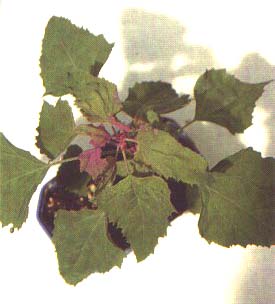
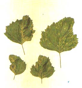
FIGURE 2 Grape leaf with typical fanleaf symptoms
FIGURE 3 Chlorotic mottle and deformation caused by fanleaf infection
FIGURE 4 Abnormal branching in a shoot of a fanleaf-infected vine
FIGURE 5 Abnormal shoot in a fanleaf-infected vine
FIGURE 7 Total yellowing of a spring shoot of a vine infected by chromogenic GFLV strain
FIGURE 9 Yellow mosaic symptoms in a leaf of an American rootstock
FIGURE 10 Spring symptoms of yellow mosaic
FIGURE 11 Totally yellow and stunted vine affected by yellow mosaic next to a normally growing plant
FIGURE 12 A patch of vines with yellow mosaic seen from a distance
FIGURE 13 Yellow mosaic symptoms fading away in late summer
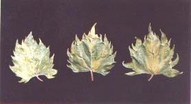
FIGURE 17 Systemic mottling induced by GFLV in Chenopodium amaranticolor
FIGURE 18 Systemic mottling and deformation of the leaves of Chenopodium quinoa infected by GFLV
FIGURE 19 Twisting of the upper leaves typically induced by GFLV infections in Gomphrena globosa
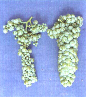
FIGURE 20 Bunches from a fanleaf-infected vine before (left) and after (right) heat therapy
Grapevine degeneration - European nepoviruses
G.P. Martelli and B. Walter
CAUSAL AGENTS
In addition to GFLV there are seven distinct nepoviruses: tomato black ring (TBRV), arabis mosaic (ArMV), raspberry ringspot (RRV), strawberry latent ringspot (SLRV), grapevine chrome mosaic (GCMV), grapevine Bulgarian latent (GBLV) and artichoke Italian latent (AILV) viruses, involved to a varying extent in the aetiology of this disease. All these viruses have isometric particles about 30 nm in diameter and the bipartite genome typical of the nepovirus group (Harrison and Murant, 1977). All possess natural serological variants, but in most cases strains infecting grapevines belong to a single serotype. Some of these viruses are serologically distantly related to one another or to other nepoviruses, i.e. GCMV to TBRV, ArMV to GFLV, GBLV to blueberry leaf mottle virus (reviewed by Martelli and Taylor, 1989). There are biological variants that elicit different symptoms in naturally and artificially infected hosts.
GEOGRAPHICAL DISTRIBUTION
European nepoviruses prevail in central and eastern Europe, where they occur in many natural and agricultural environments together with their vectors. In grapevines, ArMV is common in certain areas of France (Charentes, Alsace) and Germany (Palatinate) and is more rarely found in Switzerland, Italy, Yugoslavia, Hungary and Bulgaria. TBRV, SLRV and RRV are largely restricted to Germany (Palatinate and Moselle), even though there are occasional records from other European (Italy, France, Portugal, Turkey) and non-European (Israel) countries. GCMV and GBLV prevail in eastern Europe (Yugoslavia, Czechoslovakia, former USSR, Hungary, Bulgaria).
ALTERNATE HOSTS
ArMV, TBRV, RRV, SLRV and AILV have a wide range of wild and cultivated alternate hosts that constitute their natural reservoirs. These hosts include annual and perennial weeds, vegetable crops, shrubs, ornamentals and fruit trees. No alternate hosts for GCMV and GBLV are known.
FIELD SYMPTOMS
Like GFLV, some European nepoviruses (e.g. ArMV and GCMV) have distorting and chromogenic strains that elicit different symptomatologies, characterized by chlorotic mottling, leaf and cane deformation or chromeyellow discolorations of the foliage (Figures 21 to 25). The symptoms shown in the field by vines affected by these viruses are very similar to, if not indistinguishable from, those of GFLV-induced degeneration.
A distinctive trait of the yellowing caused by GCMV is that, unlike GFLV yellowing, it shows up equally well in the field and greenhouse. Furthermore, vines infected by GCMV lack vigour, bear little or no crop and tend to decline and die within a few years after infection. Heavy yield losses (up to 80 percent of the crop) are associated with infections by other European nepoviruses such as SLRV, TBRV and RRV (Ruder, 1985). These viruses induce leaf deformity and yellowing (Figures 26 and 27). In the cultivars Gruner Sylvaner and Riesling, SLRV elicits a most peculiar reddish discoloration of the tip of the spring shoots (Figure 28). This discoloration disappears in summer, when the vines exhibit a more or less normal appearance but are virtually fruitless (Figure 29).
NATURAL SPREAD
Experimental evidence for nematode transmission from grape to grape has been obtained only for ArMV/Xiphinema diversicaudatum and TBRV/Longidorus attenuantus. The ecology of these viruses in vineyards follows the same pattern known for other crops, i.e. seed transmission in weeds and persistence of the pathogens in their seedlings, which constitute sources of inoculum and food for the vector (reviewed by Martelli, 1978).
Although there is plenty of visual evidence that RRV and GCMV spread naturally in vineyards in a manner suggestive of soil transmission, their vectors have not been identified. Long-distance spread is through infected propagative material (budwood, rootstock roofings, grafted vines).
Rooted plants are high-risk materials because they can carry viruses and vectors together and thus establish active infection foci in any new environment favourable to active multiplication of the nematode vectors.
DETECTION
The presence of the disease in vineyards is disclosed by its symptoms. Plants can be inspected for symptoms throughout the vegetating season, but special attention should be paid to the spring growth, which ordinarily expresses the strongest reactions. Infections by mild virus strains or tolerance in certain cultivars can result in attenuated forms of disease that may escape observation. In the field it is virtually impossible to establish whether any given symptomatology is induced by one virus or another, or by a mixed infection of two or more viruses.
Sorting and identification of viruses can only be done by biological assays and/or laboratory tests.
IDENTIFICATION
Indexing by graft transmission
Any of the current graft inoculation procedures described in Part 11 can be used.
Graft transmission of GCMV to Vitis species yields foliar distortions (Figure 30) and yellow discolorations (Figure 31 ) like those induced by other nepoviruses (GFLV in particular). Somewhat more specific reactions are given by the cultivars Pinot noir and Jubileum 75, both of which show extremely severe stunting and necrosis of the apex (Figure 32). No symptoms are produced in Vitis rupestris St George, which differentiates GCMV from GFLV. In field indexing, symptoms develop on the second year's growth (Lehoczky, 1985).
Siegfriedrebe (FS4 201-39) is regarded in Germany as the best indicator for ArMV, RRV and TBRV. Cane deformations and foliar discolorations appear a few weeks after inoculation, but these symptoms are not distinctive enough to separate viruses. The Pinot noir cultivar can also be used as an indicator for TBRV.
Transmission to herbaceous hosts
All European nepoviruses are readily transmitted to
herbaceous hosts by sap inoculation. Sources and preparation of
inoculum and inoculation procedures are the same as described for
GFLV.
The separation of viruses based on differential host range responses is not easy and may pose problems even to experienced workers. Certain hosts, however, give reactions that provide useful hints for identification.
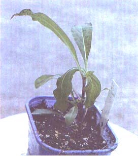
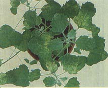
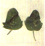
Symptoms in Chenopodium quinoa are generally milder for ArMV than for GFLV. They consist of systemic mottling and mild deformations of the leaves, with rare local lesions (Figure 33). ArMV can also induce striking chlorotic ringspotting in Nicotiana glutinosa (Figure 34).
TBRV is very severe in C. quinoa, producing distinct chlorotic/necrotic local lesions followed by systemic spread and necrosis of the plant tips (Figure 35). RRV induces local chlorotic or necrotic spots in Nicotiana rustica, followed by chlorotic/necrotic systemic rings, spots and line patterns.
SLRV may elicit chlorotic local lesions in inoculated Cucumis sativus cotyledons, followed by systemic interveinal chlorosis.
GCMV has fairly narrow host range (Martelli and Quacquarelli, 1972). Diagnostic hosts are:
GBLV also has a narrow host range (Martelli et al., 1977). Diagnostic hosts are:
Serology
Immunodiffusion tests may be used successfully with extracts from GBLV-infected grapevine leaves, especially when collected from greenhouse-grown cuttings. The same does not apply to tissue extracts of vines infected by any other European nepovirus. However, with these other viruses positive reactions are obtained using crude sap expressed directly, without addition of buffered solutions, from leaves of systemically invaded herbaceous hosts. Young, upper symptomatic leaves are the best source of antigen.
ELISA. Immunoenzymatic procedures carried out as described in Part III can be employed for the detection and identification of different European nepoviruses in naturally infected vines (Tanne, 1980; Stellmach, 1985; Walter et al., 1984; Kölber et al., 1985). Sources of antigen can be buds, roots, leaves, bark and wood scrapings. The weight of tissues used varies from 200 mg to 1 g, and the extraction buffer, as discussed for GFLV, may or may not contain nicotine ( 1 to 2.5 percent). As with GFLV, the use of buds from dormant canes or wood shavings may bypass the problem posed by the seasonal variation of virus titre, which limits the utilization of ELISA to certain growth periods (Ruder, Alebrand and Altmayer, 1983; Kölber et al., 1985). Single as well as mixed infections can be picked up by ELISA, and the viruses can be identified. Precautions for positive and negative controls are the same as with GFLV.
Immune electron microscopy (ISEM). Detection and identification of European nepoviruses by ISEM can be carried out, as outlined in Part 111, using upper symptomatic leaves collected in spring (Russo, Martelli and Savino, 1980).
Molecular hybridization
Aprobe to ArMV has been prepared (Steinkellner et al., 1989). cDNA probes to GFLV RNA- 1 or RNA-2 can also detect some ArMV isolates.
SANITATION
The same techniques used for GFLV are applicable.
REFERENCES
Bercks, R. & Stellmach, G. 1966. Nachweis verschiedener Viren in Reisigkrankheiten Reben. Phytopathol. Z., 65: 288-296.
Harrison, B.D. & Murant, A.F. 1977. Nepovirus group. Descriptions of Plant Viruses, No. 185. Kew, UK, Commonw. Mycol. Inst./Assoc. Appl. Biol.
Kölber, M., Beczner, L., Pacsa, S. & Lehoczky, J. 1985. Detection of grapevine chrome mosaic virus in field-grown vines by ELISA. Phytopathol. Mediterr., 24: 135- 140.
Lehoczky, J. 1985. Detection of grapevine chrome mosaic virus in naturally infected vines by indexing. Phytopathol. Mediterr., 24: 129-134. Martelli, G.P. 197X. Nematode-borne viruses of grapevine, their epidemiology and control. Nematol. Mediterr., 6: 127.
Martelli, G.P., Gallitelli, A., Abracheva, P., Savino, V. & Quacquarelli, A. 1977. Some properties of grapevine Bulgarian latent virus. Ann. Appl. Biol., 85: 51-58.
Martelli, G.P., Lehoczky, J. & Quacquarelli, A. 1966. Host range and properties of a virus associated with Hungarian grapevines showing macroscopic symptoms of fanleaf and yellow mosaic. Proc. Int. Conf: Virus Vector Perennial Hosts and Vitis, 1965, p. 389-401. Div. Agric. Sci., Univ. Calif., Davis.
Martelli, G.P. & Quacquarelli, A. 1972. Grapevine chrome mosaic virus. Descriptions of Plant Viruses, No. 103. Kew, UK, Commonw. Mycol. Inst./Assoc. Appl. Biol.
Martelli, G.P. & Taylor, C.E. 1989. Distribution of viruses and their nematode vectors. Adv. Dis. VectorRes., 6: 151-189.
Rudel, M. 1985. Grapevine damage induced by particular virus-vector combinations. Phytopathol. Mediterr., 24: 183- 185.
Rudel, M., Alebrand, M. & Altmayer, B. 1983. Untersuchungen Ober den Einsatz des ELISA Test zum Nachweis verschiedener Rebenviren. Vein Wiss., 38: 177-185.
Russo, M., Martelli, G.P. & Savino, V. 1980. Immunosorbent electron microscopy for detecting saptransmissible viruses of grapevine. Proc. 7th Meet. /CGV, Niagara Falls, NY, USA, 1980, p. 251 -257.
Steinkellner, H., Himmler, G., Laimer, M., Mattenovich, D., Bisztroy, G. & Katinger, H. 1989. Konstruktion von cDNA von Arabis Mosaik Virus und deren Anwendung für Diagnose. Mitt. Klosterneuburg Rebe Wein Obstbau Frht.' 39: 242246.
Stellmach, G. 1985. ELISA testing of grapevine roofings reared from nepovirus-infected mother plants forced to rapid growth. Phytopathol. Mediterr., 24: 123- 124.
Tanne, E. 1980. The use of ELISA for the detection of some nepoviruses in grapevines. Proc. 7th Meet. ICVC;, Niagara Falls, NY, USA, 1980, p. 293-296.
Walter, B., Vuittenez, A., Kuszala, A., Stocky, G., Burckard, J. & Van Regenmortel, M.H.V. 1984. Detection sérologique du virus du courtnoué de la vigne par le test ELISA. Agronomie, 4: 527-534.
Summary: grapevine degeneration detection
GRAFT TRANSMISSION
Indicators
Several Vitis vinifera cultivars: Pinot noir, Jubileum
75 (GCMV); Siegfriedrebe (ArMV, RRV, TBRV)
No. plants/test
3-5 rooted cuttings Inoculum
Wood chips, single buds, bud sticks, shoot tips
Temperature
22-24°C
Symptoms
Severe stunting and necrosis of the apex of Pinot noir in the
second year of vegetation (GCMV); foliar discolorations and cane
deformations of Siegfriedrebe within the first year after
inoculation
TRANSMISSION TO HERBACEOUS HOSTS
Diagnostic hosts
Datura stramonium (GCMV), Chenopodium quinoa (TBRV,
GBLV), Nicotiana clevelandii (RRV), Cucumis sativus (SLRV),
Nicotiana glutinosa (ArMV)
Inoculum
Tissue from young symptomatic leaves
Extraction
Grind in 2.5 percent aqueous nicotine
Temperature
Below 25°C
Symptoms
In D. stramonium (CGMV), transient, systemic,
yellowish zonate spots;
In C. quinea (TBRV), necrotic local lesions in 6-8 days,
followed by mosaic and necrosis of the plant tip in about 2
weeks;
In C. quinoa (GBLV), necrotic local lesions in 3-4 days,
systemic chlorotic mottle and necrosis;
In M clevelandii (RRV), necrotic local spots and rings in 5-7
days and systemic veinal necrosis;
In C. sativus (SLRV), chlorotic local lesions in 5-7 days
and systemic interveinal chlorosisor necrosis in 10-12 days;
In N. glutinosa (ArMV), chlorotic ringspots
OTHER TESTS
Serology (ELISA, ISEM)
Molecular hybridization
FIGURE 22 Yellow mottling induced by a chromogenic strain of ArMV
FIGURE 23 Chlorotic mottling and leaf deformation induced by a distorting strain of GCMV
FIGURE 24 Typical yellow discoloration caused by a chromogenic strain of GCMV
FIGURE 25 A row of vines with severe chrome mosaic symptoms
FIGURE 26 Yellowing and marginal necrosis in vine infected by RRV (Photo: M. Rudel)
FIGURE 27 Leaf deformity and yellowing in a vine infected by TBRV (Photo: M. Rudel)
FIGURE 31 Chrome mosaic symptoms in the indicator Kober 5BB I Photo: .1 Lehoczky)
FIGURE 33 Mosaic mottle induced by ArMV in C. quinoa
FIGURE 34 Yellow spots and rings induced by ArMV in M glutinosa
FIGURE 35 Necrotic local lesions and top necrosis induced by TBRV in C. quinoa
FIGURE 36 Chlorotic local lesions induced by GCMV in C. guinoa
FIGURE 37 Severe systemic yellowing induced by GCMV in C. guinoa
FIGURE 38 Chlorotic local lesions induced by SLRV in C. quinoa