![]()
![]()
![]()
Livestock. The fate of tebufenozide fed to lactating dairy goats was studied by dosing three goats orally with tebufenozide labelled in all three parts of the molecule at the equivalent of approximately 50 ppm in the feed for 7 consecutive days. The goats weighed between 45 and 60 kg. Milk, urine, and faeces were collected during the dosing period and the animals were killed within 24 hours after the last dose. No adverse effects of the dosing were observed. The samples collected for analysis were composite fat, consisting of equal portions of omental and perirenal fat, kidneys, liver, and composite muscle consisting of equal portions of longissimus dorsi, semimembranosus, and triceps muscle (Bender et al., 1995). The total radioactivity recovered in the excreta, tissues, and milk was 88.2% of the A-ring dose, 87.3% of the B-ring, and 90.0% of the t-butyl. Table 2 shows the recovery of the administered activity in the separate samples.
Table 2. Recovery of ingested radioactivity from goats fed with [14C]tebufenozide (Bender et al., 1995).
|
Sample |
% of total dose of 14C |
||
|
A-ring 14C |
B-ring 14C |
t-butyl 14C |
|
|
Faeces |
79.0 |
78.3 |
81.1 |
|
Urine |
8.9 |
8.6 |
7.8 |
|
Liver |
0.07 |
0.12 |
0.4 |
|
Composite fat |
0.14 |
0.15 |
0.26 |
|
Composite milk |
0.09 |
0.08 |
0.26 |
|
Composite muscle |
0.02 |
0.06 |
0.16 |
|
Kidney |
<0.01 |
<0.01 |
0.01 |
|
Heart |
<0.01 |
<0.01 |
<0.01 |
|
Total |
88.2 |
87.3 |
90.0 |
The test compound was eliminated mainly in the faeces where 79.0%, 78.3% and 81.1% of the total dose was recovered from the A-ring, B-ring, and t-butyl labels respectively. The urine accounted for 8.9% of the A-ring, 8.6% of the B-ring, and 7.8% of the t-butyl activity. Thus 87% to 89% of the administered dose was eliminated from the body via the excreta. Less than 0.3% of the dose was excreted in the milk during the 7-day dosing period: 0.09%, 0.08%, and 0.26% of the total dose from the A-ring, B-ring, and t-butyl labels respectively. Body tissues contained small amounts of activity: fat contained the highest percentage of the dose in the A-ring and B-ring samples, 0.14% and 0.15%, and the liver the highest amount from the t-butyl label (0.4% of the dose). Detectable residues were found in the muscle, heart, and kidney of all the goats. Residue levels in the tissues expressed as tebufenozide equivalents are shown in Table 3.
Table 3. Residues of 14C as tebufenozide equivalents in the milk and tissues of goats (Bender et al., 1995).
|
Sample |
14C, mg/kg tebufenozide equivalents |
||
|
A-ring 14C |
B-ring 14C |
t-butyl 14C |
|
|
Liver |
0.50 |
0.99 |
2.87 |
|
Fat |
0.17 |
0.14 |
0.29 |
|
Kidney |
0.04 |
0.06 |
0.30 |
|
Muscle |
0.007 |
0.02 |
0.06 |
|
Milk (day 2) |
0.07 |
0.07 |
. 0.16 |
Milk, fat, liver, kidney, muscle, and urine samples were analysed to determine the nature of the residue. Milk samples were extracted with chloroform, methanol and water, fat samples with hexane and methanol, and muscle, kidney and liver with various solvent mixtures which included acetonitrile, water, chloroform and methanol. Urine samples were extracted with ethyl acetate and butanol. The quantitative determination of the metabolites was carried out by TLC, GLC, HPLC and MS.
Residue levels of 14C as tebufenozide in the milk remained relatively constant throughout the dosing period. With the exception of a single residue of 0.12 mg/kg seen on day 6 from the B-ring label, residues in the A- and B-ring milk samples remained consistently in the range 0.05-0.07 mg/kg throughout the 7 days. Residues in the t-butyl milk were approximately twice those found in the other samples, from 0.09 mg/kg on day 1 to a maximum of 0.17 mg/kg on day 5.
The total radioactive residue (TRR) in the milk samples from day 2 corresponded to 0.07 mg/kg from the A- and B-ring labels and 0.16 mg/kg from the t-butyl label. More than 90% of the A- and B-ring activity was extractable with, or partitioned into, organic solvents. Only slightly more than 40% of the original t-butyl activity could be extracted in the same manner: most of the remainder, 37% of the TRR, remained in the aqueous fraction and was characterized as arising from polar, nonvolatile small molecules, probably lactose and/or amino acids. The t-butyl label also had the highest proportion of unextractable activity, nearly 10%. Hydrolysis with dilute acid or base released about one third of this activity, all of which was insoluble in ethyl acetate.
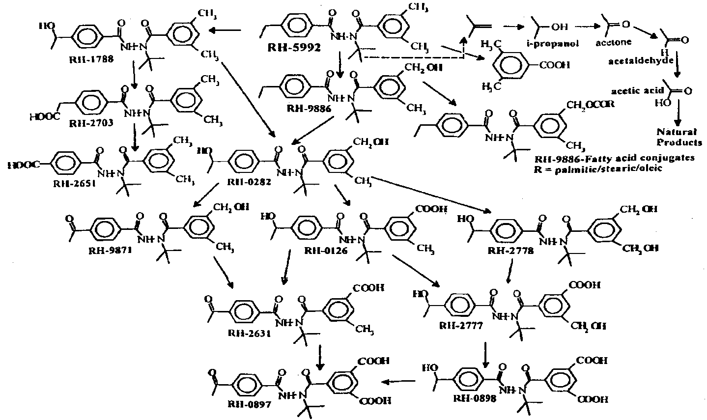
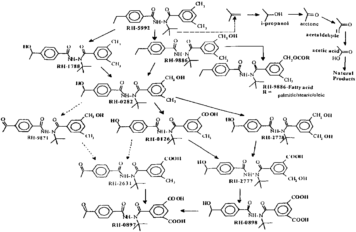
In the milk, all 3 labels appeared in the same residues, generally in similar concentrations. The milk contained two major components of the TRR. One or more fatty acid conjugates of the B-ring alcohol RH-9886 represented 17-24% of the total activity from the A- and B-ring labels (0.01-0.017 mg/kg). Tebufenozide was detected at a concentration of approximately 0.01 mg/kg from each of the labels, and represented 13.7% of the TRR. Three other alcohol metabolites were also identified: RH-0282, RH-9871 and RH-9886.
Residue levels of 14C as tebufenozide in fat samples ranged from 0.143 mg/kg from the B-ring label to 0.286 mg/kg from the t-butyl. More than 90% of the activity in the A- and B-ring labelled fat samples was extractable with or partitioned into organic solvents, but only about 55% of the t-butyl activity was in these fractions. Almost 18% of the t-butyl activity was found in the aqueous fraction, with close to 27% of the activity remaining in the post-extraction solids (PES). After saponification, most of the PES activity was released and partitioned into both aqueous and organic extracts. Radioactive residues in the fat comprised solely the parent compound and three fatty acid conjugates of RH-9886, the conjugated alcohol present in milk. The fatty acids were identified by mass spectrometry as palmitic, oleic, and stearic. The relative amounts of the parent and the conjugates varied in the three samples. Residues of tebufenozide were highest in the B-ring labelled fat, contributing 69.1% of the total residue at a concentration of 0.1 mg/kg. The lowest concentration of the parent, <0.02 mg/kg, was found with the t-butyl label, where it represented only 5.4% of the TRR.
The liver contained the highest concentration of residues with all 3 labels of any of the tissues analysed, ranging from 0.5 mg/kg from the A-ring to 2.9 mg/kg from the t-butyl label. The 3-6-fold higher residue levels in the t-butyl sample suggested some fragmentation of the parent molecule.
The major component of the A- and B-ring labelled liver was RH-2703, in which the terminal group of the ethyl substituent on the A-ring has been oxidized to carboxyl. This compound was also present in large quantities in the t-butyl labelled liver, but that also showed an approximately equal amount of one of the fatty acid conjugates of RH-9886. Numerous oxidative degradation products were present with all 3 labels. The major metabolites identified with the t-butyl label were volatile: 2-propanol and acetaldehyde. No residues of the parent were seen in the liver.
Residues in kidney samples were identified as a small amount of unmetabolized parent, together with several of its metabolites. Residues of tebufenozide were well below 0.01 mg/kg with each of the labels and represented between 0.9 and 12.8% of the TRR. Residues of RH-9886, RH-0282, and RH-2631 were also identified. The highest concentration of any single metabolite was 0.015 mg/kg of RH-0282 in the B-ring labelled sample (23.3% of the TRR in the kidney).
Muscle contained the lowest levels of radioactive residues of any samples analysed. The TRR ranged from less than 0.01 mg/kg with the A-ring label to 0.06 mg/kg with the t-butyl label. The parent and 3 of its metabolites were identified with all three labels. Samples with the B-ring label contained the highest amount of unmetabolized parent, 45.1% of the TRR, at a concentration of 0.008 mg/kg. The concentrations of tebufenozide with the A-ring and t-butyl labels were 0.002 and 0.005 mg/kg respectively (32.6% and 8.9% of the TRR). Three alcohol metabolites were detected: RH-9886, RH-0282, and RH-2778. All were present at concentrations below 0.004 mg/kg. The distribution of the identified residues is shown in Table 4.
Table 4. Residues identified in goat milk and tissues (Bender et al, 1995).
| Compound | % of total 14C in sample1 | |||||
|
Milk (day 2) |
Fat |
Liver |
Kidney |
Muscle |
||
|
Tebufenozide |
13.7 |
47.4 |
- |
11.4 |
38.9 |
|
|
Fatty acid conjugates(s) of RH-9886 |
20.8 |
20.5 |
5.0 |
- |
- |
|
|
RH-0282 |
9.3 |
- |
2.7 |
15.7 |
24.8 |
|
|
RH-9871 |
6.7 |
- |
1.2 |
- |
- |
|
|
RH-9886 |
1.0 |
- |
2.4 |
12.1 |
10.1 |
|
|
RH-2703 |
- |
- |
47.4 |
- |
- |
|
|
RH-2777 |
- |
- |
1.6 |
- |
- |
|
|
RH-2778 |
- |
- |
- |
- |
13.9 |
|
|
RH-0126 |
- |
- |
3.4 |
- |
- |
|
|
RH-2631 |
- |
- |
- |
15.0 |
- |
|
|
2-propanol |
- |
- |
44.3 |
- |
- |
|
| Acetaldehyde | - | 5.5 | - | - | ||
1 Mean percentage of A- and B-ring labels in milk, fat, kidney and muscle, and in liver for RH-0282, 9886, 2703 and 0126. Percentage of t-butyl label in liver for other compounds.
Tebufenozide is extensively metabolized by multiple oxidative transformations in goats. In most samples the parent compound represented only a small proportion of the TRR and the same residues were found with all 3 labels, indicating that the metabolites contained the intact molecular skeleton of the parent molecule. One of the resulting alcohols, RH-9886, was found conjugated to fatty acids in milk and fat. Low levels of two other alcohol metabolites and a carboxylic acid were also found in milk. Liver was exceptional in that residues of the parent compound were not found: the major metabolites identified with the t-butyl label were 2-propanol and an approximately equal amount of RH-2703. The proposed metabolic pathways of tebufenozide in lactating goats are shown in Figure 2.
Poultry. Laying hens (6 groups of 10 hens each, 25 weeks old) were dosed by capsule with labelled tebufenozide for 7 days at a level equivalent to 30 ppm in the feed. The average feed intake was 110 g/bird/day. Another group of 10 hens served as the control. Two of the 6 groups received the A-ring label, another two the B-ring and the remaining two the t-butyl label. Samples of excreta and eggs were collected daily for the 7-day dosing period, and tissues samples were collected after all the birds were killed 24 hours after the final dose. The recovery of the 14C in the A- and B-label groups was nearly quantitative, but in the t-butyl group only about 80%, probably owing to extensive degradation to produce the volatile metabolites identified in liver and possibly loss as CO2. The total radioactivity in the tissues varied with the label, and was generally highest with the t-butyl. Significantly different TRRs from the three labels implied extensive breakdown of the molecule. The average residues found in tissues and eggs are shown in Tables 5 and 6 (Schuck and Sharma, 1996).
Table 5. 14C residues in hen tissues (Schuck and Sharma, 1996).
|
Sample |
14C, mg/kg as tebufenozide, mean of 2 groups |
||
|
A-ring label |
B-ring label |
t-butyl label |
|
|
Liver |
0.13 |
0.18 |
3.95 |
|
Fat |
0.13 |
0.06 |
0.16 |
|
Thigh muscle |
0.02 |
0.01 |
0.07 |
|
Breast muscle |
0.006 |
0.0000 |
0.03 |
|
Kidney |
0.13 |
0.12 |
0.97 |
Figure 2. Proposed metabolic pathways of tebufenozide in goats.
Table 6. 14C residues in whole eggs.
|
Label |
14C, mg/kg as tebufenozide, mean of 2 groups |
||||||
|
Day 1 |
Day 2 |
Day 3 |
Day 4 |
Day 5 |
Day 6 |
Day 7 |
|
|
A-ring |
0 |
0.02 |
0.03 |
0.04 |
0.06 |
0.06 |
0.07 |
|
B-ring |
0 |
0 |
0.01 |
0.04 |
0.02 |
0.03 |
0.03 |
|
t-butyl |
0 |
0.03 |
0.05 |
0.04 |
0.09 |
0.11 |
0.13 |
The hens dosed with the t-butyl label showed the highest TRR in eggs in almost all the samples collected.
The analysis of eggs showed only traces of whole-molecule metabolites such as the parent compound, RH-9886, RH-9871 and a fatty acid conjugate of RH-9886. The analysis of fat also showed a high percentage of radiocarbon from the A-ring and t-butyl labels incorporated into the fatty acids themselves.
The t-butyl labelled liver sample contained a very high percentage of the TRR as a volatile residue. More than 30% was shown to consist of two volatile components, 2-propanol and acetaldehyde, and another volatile residue was identified as acetic acid. RH-2277, RH-2778, RH-0897, RH-0126 and RH-0282 were detected in small quantities.
The residue in muscle was very low. Solvent-extractable residues in thigh muscle amounted to about 0.03 mg/kg or less and the remainder was extractable after treatment with proteolytic enzymes.
Excreta were the main source of metabolites in this study: most of the residue was soluble in organic solvents and only a small proportion was water-soluble. The compounds were identified by TLC and HPLC, and further confirmed by mass spectrometry. Tebufenozide, RH-0126, RH-0282, RH-2777 and RH-2778 were present in amounts exceeding 10%. RH-1788, RH-9886, RH-9871, RH-0897 and 3,5-dimethylbenzoic acid were also detected.
Table 7. Metabolites identified in the eggs, tissues and excreta of hens.
|
Compound |
Residues, mg/kg |
||||
|
Eggs1 |
Liver |
Fat |
Muscle |
Excreta |
|
|
Tebufenozide |
0.005 |
|
0.18 |
nd |
Detected |
|
RH-9886 |
0.0012 |
|
|
|
Detected |
|
RH-0282 |
0.003 |
Detected |
0.03 |
0.008 |
Detected |
|
RH-9871 |
0.003 |
|
|
|
Detected |
|
RH-9886 conj. |
0.0023 |
|
0.01 |
|
|
|
RH-1788/9886 |
|
|
|
0.0053 |
Detected |
|
RH-2631 |
|
|
|
0.0044 |
|
|
RH-2778 |
|
Detected |
|
<LOD |
Detected |
|
Rh-0126 |
|
Detected |
|
<LOD |
Detected |
|
RH-0897 |
|
Detected |
|
|
|
|
RH-2777 |
|
Detected |
|
<LOD |
Detected |
|
3.5-dimethylbenzoic acid |
|
|
|
|
Detected |
|
acetaldehyde + 2-propanol |
|
2.56 |
|
|
Detected |
|
acetic acid |
|
0.45 |
|
|
|
|
others & unknown |
0.045 |
|
0.013 |
|
|
|
Natural product incorporation |
>20% |
|
>30% |
|
|
1 A-ring label groups collected on day 5, B-ring and t-butyl label groups on day 7
2 A-ring label only
3 A-ring and t-butyl label
4 A- and B-ring labels
5 t-butyl label only
The metabolic degradation of tebufenozide in hens proceeded via oxidation of the ethyl and methyl groups of the A- and B- rings, hydrolysis of the amide portions which released the free benzoic acids and oxidative degradation of the tert-butyl group which resulted in the small volatile molecules 2-propanol, acetaldehyde and acetic acid. A proposed degradation pathway in hens is shown in Figure 3.
Fish. To study the kinetics of the uptake and elimination of tebufenozide, Bluegill sunfish were continuously exposed to a nominal concentration of 50 mg/l for 29 days. Each of three groups was exposed to [14C] tebufenozide with one of the three labels. Thirty-five fish were then transferred from each of the three exposure aquaria to their respective depuration aquaria for a 15-day depuration period in fresh water, to determine the half-life for the loss of tebufenozide from tissues. Fish, divided into edible and inedible components, and water samples were taken at eight intervals during the exposure period and at 5-day intervals during the depuration phase, and analysed by radioassay (Christensen, 1992).
The concentrations of 14C in the edible and inedible tissues and whole bodies of the fish reached a statistically determined steady state during the first day of exposure. The mean steady state concentration in the tissues and the bioconcentration factor (BCF) for each of the three radiolabels is shown below.
|
Label |
14C, mg/kg as tebufenozide, and bioconcentration factors in fish |
|||||
|
Edible tissue |
Inedible tissue |
Whole body |
||||
|
mg/kg |
BCF |
mg/kg |
BCF |
mg/kg |
BCF |
|
|
A-ring |
0.46 |
8.7 |
4.30 |
81 |
2.20 |
42 |
|
B-ring |
0.32 |
5.9 |
8.3 |
150 |
3.80 |
70 |
|
t-butyl |
0.41 |
8.0 |
4.5 |
88 |
2.20 |
43 |
Figure 3. Proposed metabolic pathways of tebufenozide in hens.
The reported half-life for depuration was less than three days for all labels. By the last (15th) day of depuration at least 90% of the radioactivity had been eliminated from the fish.
In another study to determine the rate and extent of tebufenozide bioconcentration in fish, Bluegill sunfish were also exposed for 29 days to nominal concentrations of 50 mg/l of tebufenozide labelled at the three sites as before. The concentration in the fish rapidly reached a steady state level. The BCF was 7.5 for the edible tissue and 52 for the whole body. Other fish were exposed to tebufenozide labelled in the A-ring at a nominal concentration of 300 mg/l for 14 days for the identification of metabolites.
The main residue in the fish extracts was unmetabolized tebufenozide but eight metabolites, RH-0126, RH-2777, RH-2778, RH-2652, RH-2631, RH-0282, RH-9886 and the A-ring ketone/B-ring diol of tebufenozide, were also isolated and identified (Dong and Hawkins, 1993).
The metabolic profiles in the edible and inedible tissues were the same: they are shown in Table 8. No other metabolites constituted more than 10% of the residue in any tissue. The proposed metabolic pathways for tebufenozide in fish are shown in Figure 4.
Figure 4. Proposed metabolic pathways of tebufenozide in fish.
Table 8. Residues in edible and inedible tissues of fish exposed to [14C]tebufenozide (Dong and Hawkins, 1993).
|
Compound |
% of total residue |
|||
|
Edible tissue |
Inedible tissue |
|||
|
t-butyl label |
B-ring label |
t-butyl label |
B-ring label |
|
|
Tebufenozide |
45.6 |
58.8 |
39.4 |
17.3 |
|
RH-0126 |
10.5 |
17.8 |
19.7 |
21.8 |
|
RH-2652 |
2.3 |
1.9 |
3.6 |
4.2 |
|
RH-2778 |
5.3 |
3.6 |
2.7 |
5.0 |
|
RH-2777 |
2.9 |
5.0 |
1.8 |
3.8 |
|
RH-0282 |
2.0 |
0.9 |
0.2 |
0.2 |
|
RH-2631 |
* |
* |
1.3 |
2.5 |
|
RH-9886 |
* |
* |
* |
* |
|
A-ring ketone/B-ring diol |
* |
* |
* |
0.2 |
* Below the limit of detection in fish exposed to 50 mg/l, but observed in fish exposed to 300 mg/l
![]()
![]()
![]()